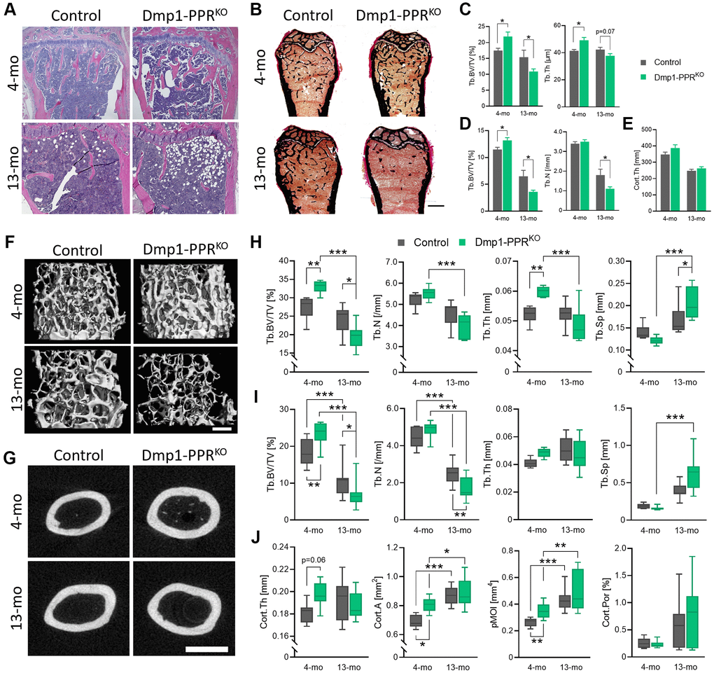
Figure 1.Age-dependent bone loss in Dmp1-PPRKO mice. Vertebrae and long bones of male control and KO animals were analyzed by (A) histology, (B–E) histomorphometry and (F–J) μCT. (A) Representative H&E of the proximal tibiae and (B) Von Kossa staining of the distal femora. Bar = 1.0 mm. Histomorphometric analysis of (C) the L5 and (D) trabecular and (E) cortical region in the distal and midshaft femora, respectively. N = 6–10 per group. Data are presented as mean ± SEM. Representative μCT images of (F) the distal and (G) the midshaft femora. Bars = (F) 300 μm and (G) 1.0 mm. μCT analysis of (H) the L5 and (I) the distal femur (trabecular) and (J) the midshaft femur (cortical) are shown. Data are presented as box and whisker plot. N = 6–16 per group. See Tables 1 and 2 for the full list of parameters. Analyses were performed in a blinded fashion. Unpaired Student’s t test (C–E) and Two-way ANOVA with Tukey’s post hoc test or Mann-Whitney test (H–J) was performed. *p < 0.05, **p < 0.01, ***p < 0.001. Abbreviations: Tb: Trabecular; Cort: cortical; BV: bone volume; TV: total tissue volume; Th: thickness; N: number; Sp: separation; A: area; pMOI: polar moment of inertia; Por: porosity.