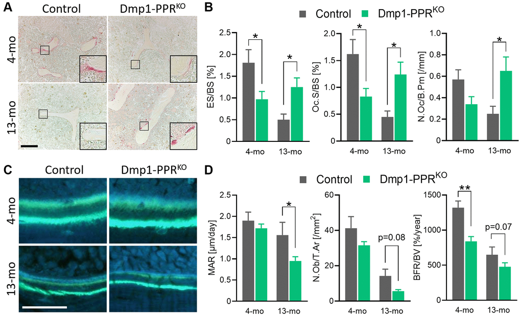
Figure 2.Histomorphometric analysis of trabecular bones of Dmp1-PPRKO mice. (A, B) Representative TRAP staining images and bone resorption parameters of the distal femora from male control and KO animals. The inset shows a closeup displaying the TRAP-positive osteoclasts on the trabecular bone surface. Bar = 200 μm. (C) Representative images of calcein double-staining on the distal femora of these mice. Bone formation within 7 days was visualized by double calcein labeling. Bar = 50 μm. (D) Representative bone-formation parameters of the distal femora are shown. See Table 1 for the full list of resorption and formation parameters. N = 6–10 per group. Analyses were performed in a blinded fashion. Unpaired Student’s t test was performed. *p < 0.05, **p < 0.01. Data are presented as mean ± SEM. Abbreviations: ES: Erosion surface; BS: bone surface; Oc.S: osteoclast surface; N.Oc: number of osteoclasts; B.Pm: bone perimeter; MAR: mineral apposition rate; N.Ob: number of osteoblasts; T.Ar: tissue area; BFR: bone formation rate; BV: bone volume.
Figure 2 — Parathyroid hormone signaling in mature osteoblasts/osteocytes protects mice from age-related bone loss | Aging