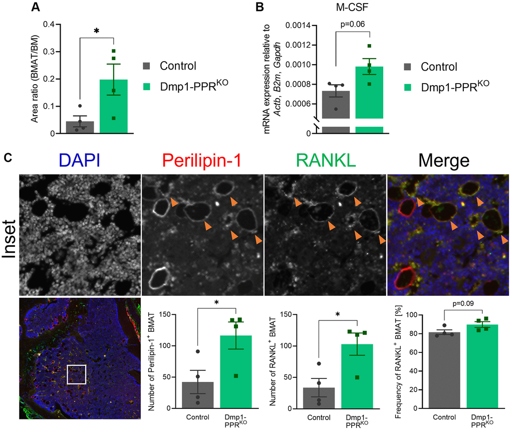
Figure 4.Increased bone marrow adipocytes in middle-aged Dmp1-PPRKO mice. (A) The area of bone marrow adipose tissue (BMAT) over the total bone marrow (BM) space within 300-μm from the epiphyseal plate was analyzed on H&E-stained tibiae sections of male control and Dmp1-PPRKO mice at 13 months of age. Representative images are shown in Figure 1A. N = 4 per group. (B) Expression of M-CSF in the BM isolated from the femora of middle-aged male animals (13 months old) was analyzed by qPCR. N = 4 per group. (C) Immunofluorescence staining of perilipin-1, RANKL and DAPI was performed on the tibiae of middle-aged (13 months) male control and Dmp1-PPRKO mice. Representative images of a tibia from Dmp1-PPRKO mouse are shown. In the merged image, DAPI, perilipin-1 and RANKL staining is shown in blue, red, and green, respectively. The orange arrowheads indicate RANKL+ BMAT (identified as perilipin-1+). The number of BMAT (left) and the number (middle) and frequency (right) of RANKL+ BMAT in the BM space were analyzed. N = 4 per group. Unpaired student’s t test was performed. *p < 0.05. Data are presented as mean ± SEM.