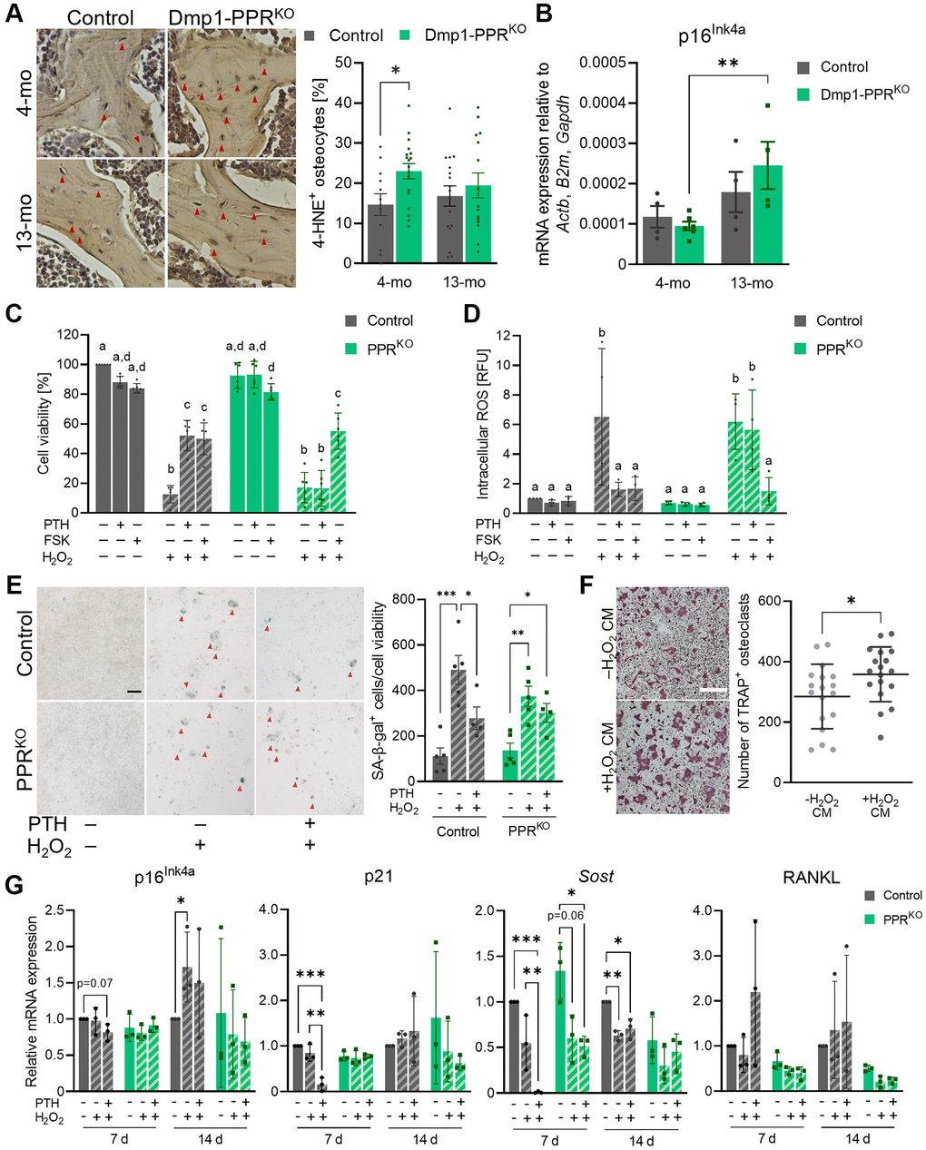
Figure 6.PTH protects osteocytes from oxidative stress-induced cell death and senescence. (A) Representative images of immunohistochemistry for 4-HNE on the L3/4 vertebrae from male animals are shown. The frequency of 4-HNE+ osteocytes per image field was analyzed. Mean ± SEM is shown. (B) Expression of p16Ink4a in the tibiae of male control and Dmp1-PPRKO mice was analyzed by qPCR. N = 4–6 per group. Mean ± SEM is shown. (C–E) Control and 12H-PPRKO osteocytic cell line was pretreated with either 10 nM hPTH(1–34) or 10 μM forskolin (FSK) for 18-22 hrs prior to H2O2 exposure. (C) After H2O2 exposure (1 mM, overnight), cell viability was measured by resazurin-based assays. (D) After H2O2 exposure (1 mM, 4 h) intracellular ROS levels were measured using a fluorescent probe (DCFDA). Data are presented as relative fluorescence unit (RFU). (E) After continuous exposure to H2O2 (150 μM, 14 d), cells were stained for SA β-gal. Representative SA β-gal staining images and the quantification of SA β-gal+ cells (red arrowheads) are shown. Bar = 100 μm. (F) Representative TRAP staining images are shown of BMMCs isolated from 3–4-month-old male control mice under osteoclastic differentiation in the presence of conditioned medium from H2O2-treated control osteocytic cell line (–H2O2 or +H2O2, 150 μM for 7 days). Bar = 400 μm. N = 18 per group. (G) After continuous exposure to H2O2 (100 or 150 μM, 7 or 14 d), cells were harvested for RNA isolation. mRNA expression of p16Ink4a, p21, Sost, and RANKL were analyzed by qPCR. N = 3 per group. Kruskal-Wallis test with Dunn’s post hoc test, two-way ANOVA with Tukey’s post hoc test, one-way ANOVA with Sidak’s post hoc test or Mann-Whitney test were performed. *p < 0.05, **p < 0.01, ***p < 0.001. Same letter indicates n.s. Data are presented as mean ± SD.