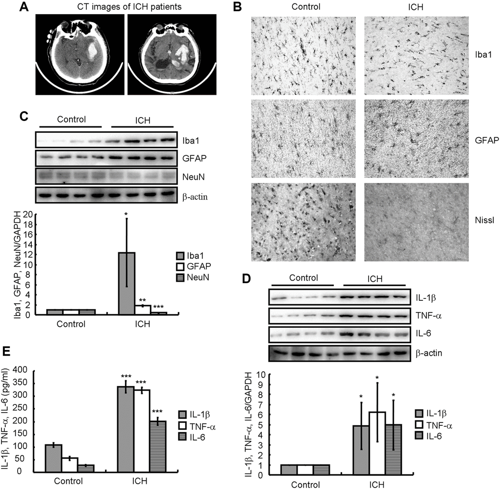
Figure 1.Neuroinflammation and neuronal loss were present in ICH patients. (A) A computed tomography (CT) image indicates hematoma formation in an ICH patient compared to a control subject. (B) Paraffin slices immunostained for Iba1 or GFAP show microglia and astrocytes, respectively. The morphology of neurons is revealed by Nissl staining. (C, D) Western blotting detects the protein expression of Iba1, GFAP, NeuN, IL-1β, TNF-α, and IL-6 in a patient with ICH. GAPDH serves as an internal control. (E) Concentrations of IL-1β, TNF-α, and IL-6 in the brain tissues of ICH patients were detected by ELISA. The results represent the mean ± SD for the repeated experiments. *P < 0.05; **P < 0.01; ***P < 0.001 vs. control subjects.