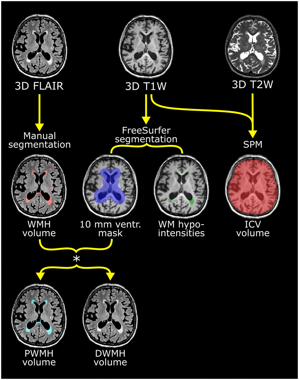
Figure 4.Flow chart of the multiparametric analysis of the brain MRI scans exemplified with scans from one participant. Upper row shows the 3D FLAIR, T1- and T2-weighted scans used. See Table 10 for scan details. Manual segmentation of white mater hyperintensities (WMH) was performed on the FLAIR images. From the T1-weighted scan, the ventricular (ventr.) mask derived with FreeSurfer (shown in violet in middle row) was used to stratify WMH into periventricular white mater hyperintensities (PWMH) (located <10 mm from the ventricular edge) and deep white mater hyperintensities (DWMH) (≥10 mm from ventricular edge) as seen in the lower row. PWMH are depicted in cyan and DWMH in white. White matter (WM)-hypointensity volume was also derived from the FreeSurfer analysis (middle row) and shown as green lesions. Intracranial volume (ICV) was calculated from the T1- and T2-weighted scans using SPM. These segmentations were performed separately for the scans from baseline, one-, three- and five years.