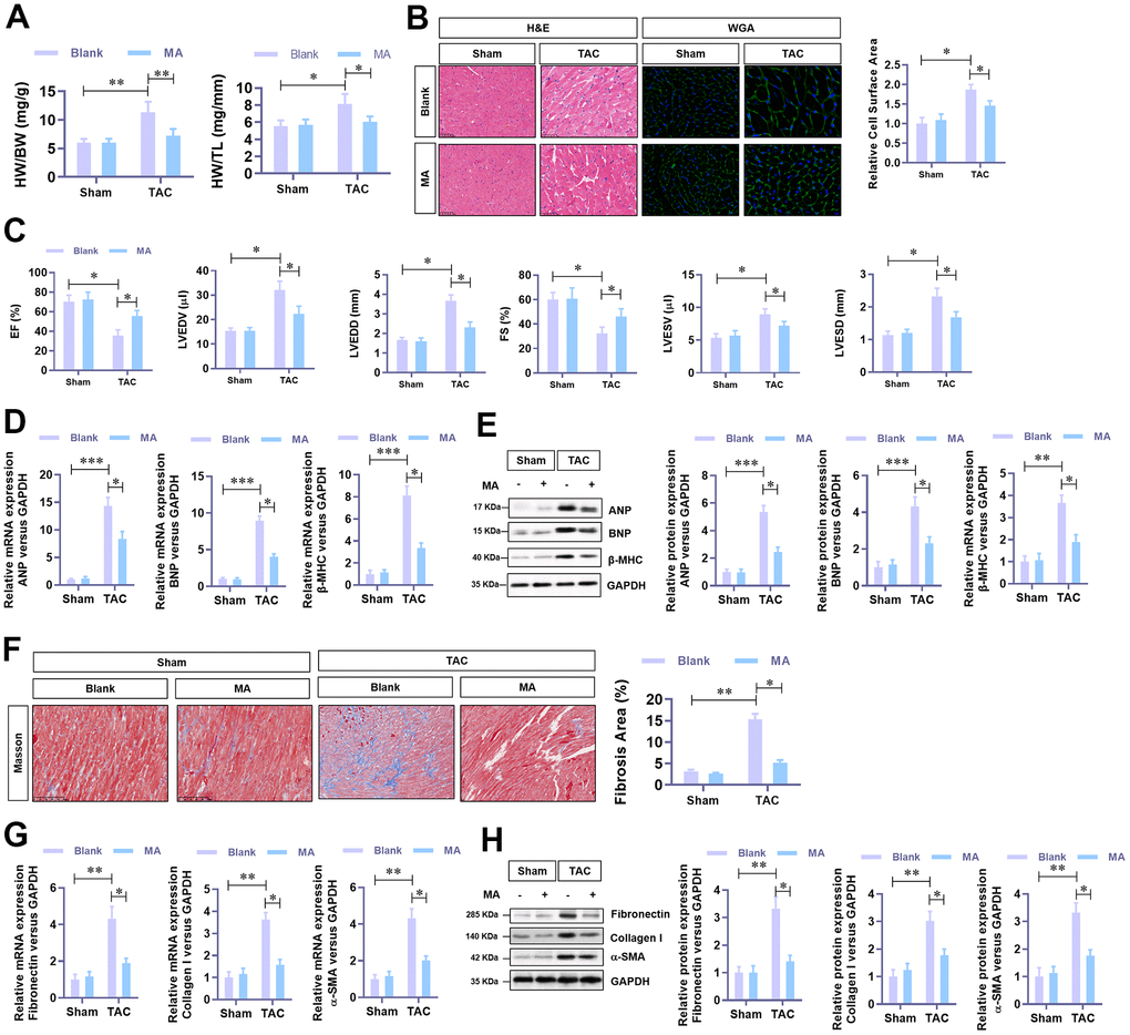
Figure 2.The protective role of MA against TAC-induced cardiac hypertrophy in vivo. (A) The ratio of HW/BW and HW/ TL, showing the effect of MA on the morphology of cardiac morphology. (B) Analysis of the effect of MA on the cardiomyocyte area in left ventricle through Histology H&E and WGA staining of cardiac cross-sections. (C) Echocardiography detection of the parameters of LVEF, LVFS, LVEDV, LVESV, LVEDD, and LVESD to evaluate the effect of MA on the cardiac function of TAC mice. (D) Real-time PCR analysis of mRNA levels of the hypertrophy markers (ANP, BNP, and β-MHC). (E) Western blot analysis of the protein levels of the hypertrophy markers (ANP, BNP, and β-MHC). (F) Masson staining detection of the effect of MA on TAC-induced myocardial fibrosis. (G) Real-time PCR analysis of mRNA levels of the myocardial fibrosis (Fibronectin, Collagen I, and α-SMA). (H) Western blot analysis of the protein levels of the myocardial fibrosis (Fibronectin, Collagen I and α-SMA); *P<0.05, **P<0.01 compared to the indicated group.