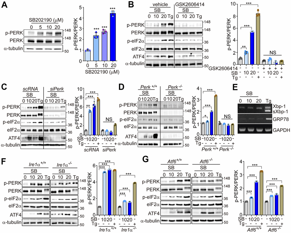
Figure 1.SB202190 activates the PERK/eIF2α/ATF4 pathway. (A) HEK293 cells were treated with SB202190 (0, 5, 10, and 20 μM) for 6 h. PERK phosphorylation was determined by western blotting. Quantification of p-PERK is shown in the right panel. (B) HEK293 cells were incubated with SB202190 (10 and 20 μM) for 6 h after pretreatment with or without the PERK inhibitor, GSK2606414 (1 μM) for 1 h. Thapsigargin (Tg, 2 μM) was used as a positive control. Cell lysates were used for western blotting analysis for p-PERK, PERK, p-eIF2α, eIF2α, and ATF4. (C) For knockdown of Perk, SH-SY5Y cells were transfected with control siRNA (scRNA) or siPerk for 48 h and then treated with different doses (10 and 20 μM) of SB202190 (SB) or Tg (2 μM) for 6 h. Cell lysates were measured for PERK activation by western blot using the indicated antibodies. (D) Perk+/+ and Perk-/- MEFs were treated with SB202190 (10 and 20 μM) for 6 h or Tg (2 μM). The levels of p-PERK, PERK, p-eIF2α, eIF2α, and ATF4 were measured by western blotting. (E) SH-SY5Y cells were treated with SB202190 (10 and 20 μM) or Tg (2 μM) for 6 h. Xbp-1 splicing and Grp78 expression were detected by RT-PCR. (F, G) Hepatocytes isolated from Ire1α +/+, Ire1α -/- (F) or Atf6α +/+, and Atf6α -/- (G) mice were treated with SB202190 (10 and 20 μM) for 6 h to assess the levels of PERK phosphorylation and ATF4 expression by western blotting. Quantification of p-PERK is shown in the right panel (A–D, F, G). Data are mean ± SD (n=3); *p<0.05, **p<0.01, and ***p<0.001.