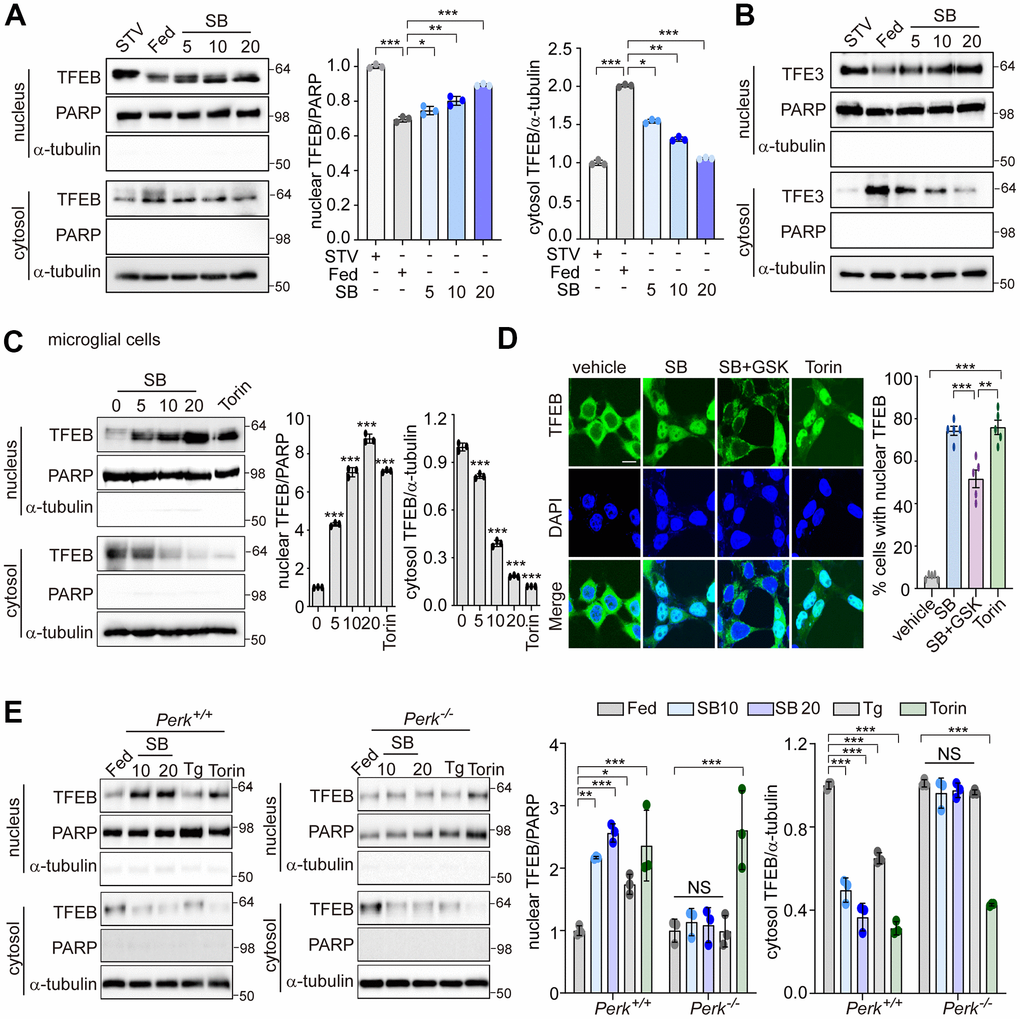
Figure 3.PERK is required for TFEB nuclear translocation by SB202190. (A, B) SH-SY5Y cells were treated with SB202190 at the indicated concentrations (5, 10, and 20 μM) for 4 h and subjected to nuclear and cytosolic fractionation. (A) Resulting fractions were then detected with antibody against TFEB. Starvation (STV) was used as positive control. PARP and α-tubulin were used as nuclear and cytosolic markers, respectively. Quantification of TFEB translocation is shown at the right. Data are mean ± SD (n=3), *p<0.05; **p<0.01; ***p<0.001. (B) The translocation of TFE3 was analyzed by western blotting. (C) The human microglial HMC3 cells were treated with SB202190 (0, 5, 10, and 20 μM) for 6 h. Torin-1 (2μM) treatment was used as a positive control. The fractionated HMC3 cells were evaluated for TFEB translocation by western blotting (left). Quantification of TFEB translocation was analyzed (right). Data are mean ± SD (n=3), ***p<0.001. (D) TFEB-EGFP-transfected SH-SY5Y cells were treated with 20 μM SB202190 in the presence or absence of the PERK inhibitor (GSK2606414, GSK). Torin-1, an mTOR inhibitor, was used as a positive control. The fluorescence of TFEB was visualized by confocal microscopy (left). Scale bar: 10 μm. Cells were evaluated to calculate the percentage of cells showing nuclear TFEB localization. n > 20 cells per condition (right). Data are mean ± SD; **p<0.01 and ***p<0.001. (E) Perk+/+ and Perk-/- MEFs were treated with SB202190 (10 and 20 μM) for 6 h or Tg (2 μM). Torin-1 (2μM) treatment was used as a positive control. Cells were detected with TFEB antibody in the nuclear and cytosol fraction by western blotting (left). Quantification of TFEB translocation was analyzed (right). Data are mean ± SD (n=3), *p<0.05; **p<0.01; ***p<0.001; NS, not significant.