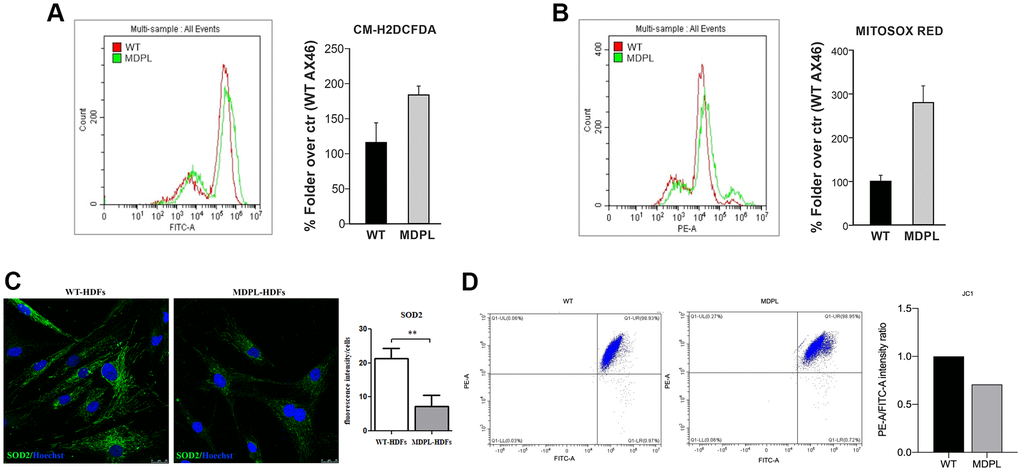
Figure 3.Functional mitochondrial evaluation of MDPL and WT HDFs. Flow cytometry quantification of (A) total reactive oxygen species using CM-H2DCFDA in HDFs WT and MDPL ones. (B) Flow cytometry quantification of mitochondrial superoxide using MitoSOX Red in HDFs WT and in MDPL ones. The error bar indicated in panels A and B is the average of two independent experiments. (C) Confocal analysis following SOD2 immunofluorescence and its quantification (**p < 0.01). (D) JC1 staining for flow cytometry to evaluate changes in mitochondrial potential membrane and the quantification of red/green fluorescence intensity ratio.