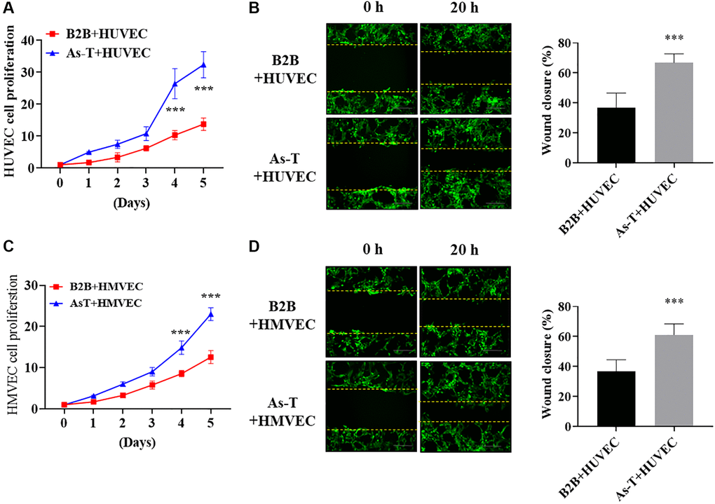
Figure 2.As-T cells increased cell migration and proliferation of human umbilical vein cells (HUVECs) and HMVECs. HUVECs or HMVECs were transduced by lentivirus expressing GFP and selected with puromycin. The GFP-positive HUVECs or HMVECs were then co-cultured with B2B or As-T cells in a 1:1 ratio. (A) The growth curve of HUVECs was plotted by counting the GFP-positive cells on indicated days. ***p < 0.001 compared to B2B+HUVEC group. (B) When reached about 100% confluence, the cells were starved overnight and a scratch wound was made with a 200 μL pipet tip. Cells were washed using PBS three times and 2 mL fresh basic EBM2 medium was added to the wells. The width of the wound was measured at 0 h and 20 h post scratch. Images were acquired at 10× magnification, bar = 100 μm. Left panel: representative images; right panel: quantification of wound healing closure from three experiments. ***p < 0.001 compared to B2B+HUVEC group. (C) The growth curve of HMVECs was plotted by counting the GFP-positive cells on indicated days. ***p < 0.001 compared to B2B+HMVEC group. (D) The wound healing assay for HMVEC cells was performed as described above. Images were acquired at 10× magnification, bar = 100 μm. ***p < 0.001 compared to B2B+HMVEC group.