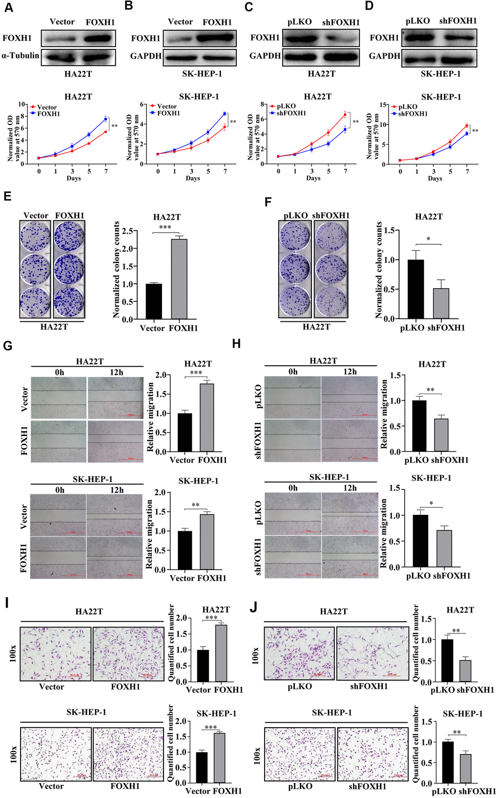
Figure 4.Experimental evidence suggested that FOXH1 promoted HCC development. (A, B) Top, the efficiency of FOXH1 overexpression. GAPDH was internal control. Bottom, MTT assay demonstrated that overexpression of FOXH1 promoted cell growth of HA22T (A) and SK-HEP-1 cells (B). (C, D) Top, efficiency of FOXH1 knockdown. GAPDH was loading control. Bottom, MTT assay revealed that knockdown of FOXH1 reduced cell growth of HA22T (C) and SK-HEP-1 cells (D). (E) FOXH1 HA22T cells had better colony forming ability than Vector HA22T cells. Left, representative images of colonies. Right, statistical analysis. (F) FOXH1 depleted HA22T cells formed less colonies than the control cells. Left, representative images of colonies. Right, statistical analysis. (G) Overexpression of FOXH1 increased cell migrating ability of HA22T (top) and SK-HEP-1 cells (bottom) cells. Left, representative images of wounding healing assay. Right, statistical analysis. (H) FOXH1 knockdown suppressed the migrating ability of HA22T (top) and SK-HEP-1 cells (bottom) cells. Left, representative images of wounding healing assay. Right, statistical analysis. (I) Overexpression of FOXH1 promoted cell invasion of HA22T (top) and SK-HEP-1 (bottom) cells. Left, representative images of wounding healing assay. Right, statistical analysis. (J) Knockdown of FOXH1 decreased cell invasion of HA22T (top) and SK-HEP-1 (bottom) cells. Left, representative images of wounding healing assay. Right, statistical analysis. *P<0.05, **P<0.01, ***P<0.001.