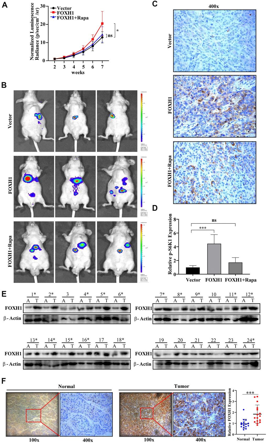
Figure 6.In vivo and clinical evidence confirmed the oncogenic role of FOXH1 in HCC development. (A) The HA22T tumor growth curve. (B) Representative images of HA22T tumors. (C) p-S6K1 staining showed that FOXH1 promoted HA22T tumor growth was dependent of mTOR activation. (D) A statistical analysis of p-S6K1 level. (E) Western blotting analysis of FOXH1 in 24 HCC samples with their paired adjacent liver tissues. (F) IHC staining of FOXH1 in HCC samples using their corresponding adjacent liver tissues as controls. Left, representative image of FOXH1 staining in slides made from patient 1#. Right, statistical analysis of FOXH1 staining. Scale bar: 25 μm. *P<0.05, **P<0.01, ***P<0.001.