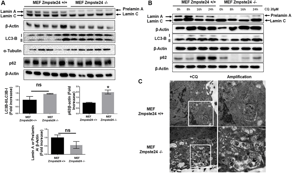
Figure 1.Immortalized MEF Zmpste24 KO cells present an increase in the basal autophagic flux. (A) Immunoblot analysis of Lamin A (in MEF Zmpste24 WT cells), prelamin A (in MEF Zmpste24 KO cells), LC3B II-I ratio and p62 using β actin as loading control, in the cell extracts in basal state (n = 5). The plot indicates the quantification data of Lamin A or prelamin A/β-actin ratio, p62/β-actin ratio and LC3B II/LC3B I ratio in the basal state. Data represent the mean ± standard error of the mean (SEM). Differences were determined by unpaired Student t-test analysis. *p < 0,05 (n = 5). (B) Immunoblot analysis of Lamin A (in MEF Zmpste24 WT cells), prelamin A (in MEF Zmpste24 KO cells), LC3B II-I ratio and p62 using β actin as loading control, in the cell extracts treated with chloroquine (CQ) (20 μM) during 0, 8, 16 and 24 hours (n = 5). (C) Electron microscopy of MEF Zmpste24 WT and KO cells treated with CQ (20 μM) 24 hours. Enlargement of areas with accumulation of autophagosomes.