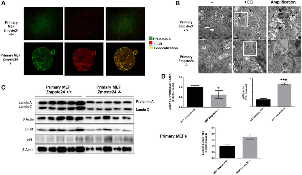
Figure 4.Primary cells from Zmpste24 KO cells also exhibit an increase in autophagic flux. (A) Immunofluorescence of Primary MEF Zmpste24 WT and KO cells under basal state using both an LC3B II/I antibody and a prelamin A antibody to see the co-localization signal. (B) Electron microscopy of Primary MEF Zmpste24 WT and KO cells in both basal states and treated with CQ (20 μM) during 24 hours. Enlargement of areas with accumulation of autophagosomes after treated with CQ. (C) Immunoblot analysis of Lamin A (in Primary MEF Zmpste24 WT cells), prelamin A (in Primary MEF Zmpste24 KO cells), LC3B II-I ratio and p62 using β actin as loading control, in the cell extracts in basal state (n = 5). (D) The plot indicates the quantification data of Lamin A or prelamin A/β-actin ratio, p62/β-actin ratio and LC3B II/LC3B I ratio in the basal state. Data represent the mean ± standard error of the mean (SEM). Differences were determined by unpaired Student t-test analysis. *p < 0,05; ***p < 0,005 (n = 5).