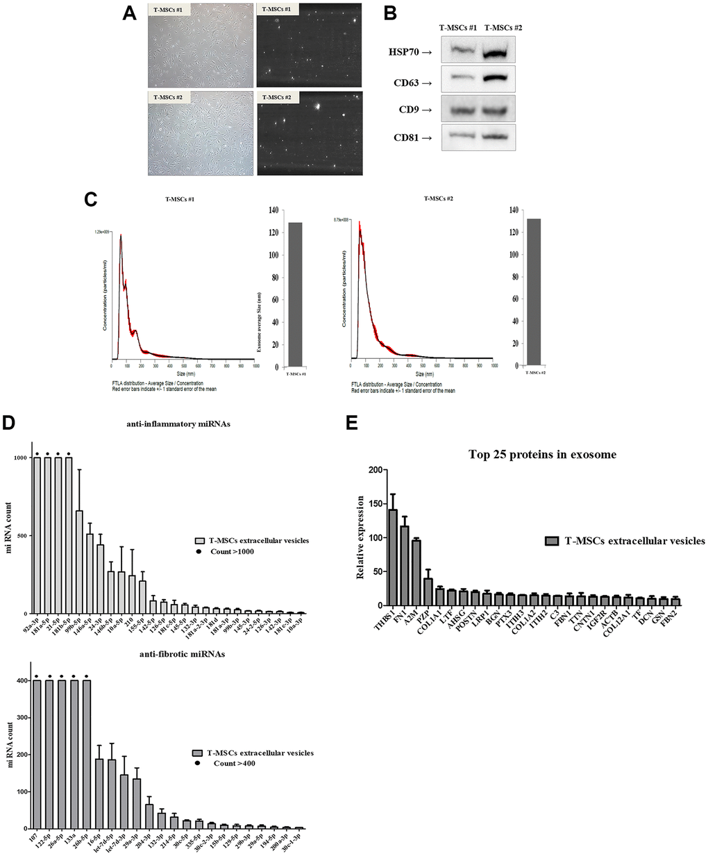
Figure 2.Identification of T-MSCs derived-extracellular vesicles. Nanoparticle tracking analysis of T-MSCs derived-extracellular vesicles. Normal cell (left) and screen shot from video of light scatter of placental vesicles overlaid with a graph of vesicle size and concentration, as determined by nanoparticle tracking analysis (right) (A). Western blot illustrating the characteristic surface markers of extracellular vesicles, HSP70, CD63, CD9, and CD81, present in the extracellular vesicles (B). Histogram of the nanoparticle tracking analysis demonstrating the size distribution of T-MSCs-derived extracellular vesicles after isolation through centrifugation (C). The presence of microRNA (miRNAs) in T-MSC-derived extracellular vesicles. T-MSCs-derived exosomes contain approximately 26 anti-inflammatory miRNAs and approximately 22 anti-fibrotic miRNAs (D). T-MSCs-derived extracellular vesicles contain 25 proteins (E).