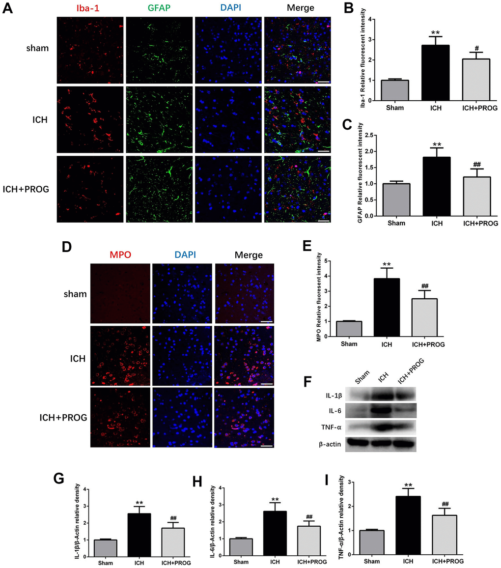
Figure 3.Effect of progesterone on the activation of microglial and astrocyte, neutrophil infiltration, neuroinflammation. (A) Representative immunofluorescence staining images of Iba-1(red) and GFAP (green) in perihematomal region. Nuclei were counterstained with DAPI (blue). Bar=50μm. (B, C) Quantitative analyses of Iba-1 and GFAP relative fluorescent intensity in perihematomal region in each group. (D) Representative immunofluorescence staining images of MPO (red) in perihematomal region. Nuclei were counterstained with DAPI (blue). Bar=50μm. (E) Quantitative analyses of MPO relative fluorescent intensity in perihematomal region in each group. (F) Representative Western bands showing the protein expression of IL-1β, IL-6, TNF-α in perihematomal region. (G–I) Quantitative analysis of Western blots shows that the expression of IL-1β, IL-6, TNF-α changes in each group. n = 6 animals per group. Data are expressed as the mean ± SEM; **P < 0.01 vs. sham; #P < 0.05 vs. ICH group; ##P < 0.01 vs. ICH group. GFAP: glial fibrillary acidic protein; Iba-1: ionized calcium binding adapter molecule 1; MPO: myeloperoxidase.