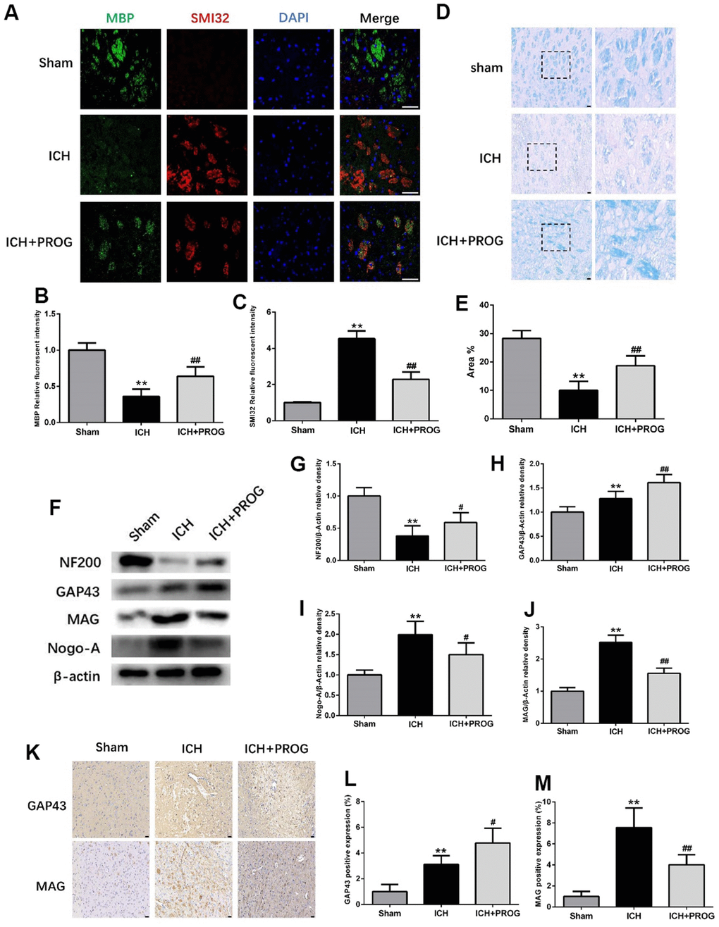
Figure 4.Effect of progesterone on myelin loss and axonal pathology. (A) Representative immunofluorescence staining images of MBP (green) and SMI32(red)in perihematomal region. Nuclei were counterstained with DAPI (blue). Bar=50μm. (B, C) Quantitative analyses of MBP and SMI32 relative fluorescent intensity in perihematomal region in each group. (D) Representative images of Luxol fast blue staining. Bar=50μm. (E) Quantitative analyses of positive stained myelin ratio in each group. (F) Representative Western bands showing the protein expression of NF200, GAP43, Nogo-A and MAG in perihematomal region. (G–J) Quantitative analysis of Western blots shows that the expression of NF200, GAP43, Nogo-A and MAG changes in each group. (K) Representative images of immunohistochemistry staining of GAP43 and MAG in perihematomal region. Bar=50μm. (L, M) Quantitative analyses of GAP43 and MAG positive expression in perihematomal region in each group. n = 6 animals per group. Data are expressed as the mean ± SEM; **P < 0.01 vs. sham; #P < 0.05 vs. ICH group; ##P < 0.01 vs. ICH group. MBP: myelin basic protein; SMI32: SMI32: Stemberger Monoclonal Incorporated Antibody 32; GAP43: Growth associated protein-43; NF200: Neurofilament; Nogo-A: Neurite outgrowth inhibitor-A; MAG: Myelin Associated Glycoprotein.