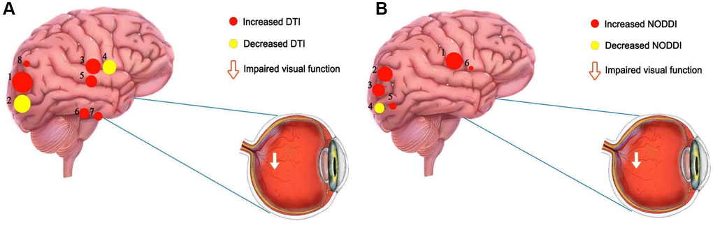
Figure 5.The mean DTI and NODOI values of altered brain regions. (A) Compared with the HCs, the DTI values of the following regions were decreased to various extents: 2- left superior longitudinal fasciculus (FA) (t = −6.78), 4- body of corpus callosum (FA) (t = −6.25). Compared with the HCs, the DTI values of the following regions were increased to various extents: 1- left superior longitudinal fasciculus (MD) (t =7.61), 7- right posterior limb of internal capsule (MD) (t = 5.31), 8- right posterior thalamic radiation (MD) (t = 4.94), 3- genu of corpus callosum (AD) (t = 6.37), 6- right posterior limb of internal capsule (AD) (t = 5.36), 5- right splenium of corpus callosum (AD) (t = 5.47). (B) Compared with the HCs, the NODOI values of the following regions were increased to various extents: 2- left anterior corona radiata (t = 5.17), 6- body of corpus callosum (t = 4.18), 5- left superior longitudinal fasciculus (ODI) (t = 4.54), 1- right splenium of corpus callosum (t = 6.86), 3- left posterior corona radiata (t = 5.03). Compared with the HCs, the NODOI values of the following regions were decreased to various extents: 4- left superior longitudinal fasciculus (FICVF) (t = −4.56). Abbreviations: HCs: healthy controls; DTI: diffusion tensor imaging; FA: fractional anisotropy; MD: mean diffusivity; AD: axial diffusivity; ODI: orientation dispersion index; FISO: isotropic volume fraction; FICVF: intracellular volume fraction.