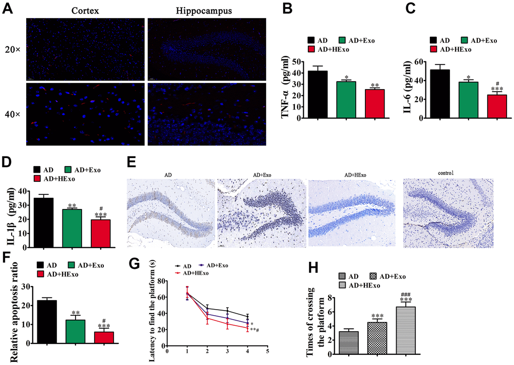
Figure 2.Exosomes from hypoxia-pretreated adipose-derived mesenchymal stem cells have a more therapeutic effect at improving cognitive function by decreasing neuronal damage in the hippocampus. (A) The 1,1′-dioctadecyl-3,3,3′,3′-tetramethylindocarbocyanine perchlorate-labeled exosomes are red and nuclei are counterstained with 4′,6-diamidino-2-phenylindole (blue). The injected exosomes were detected in the cortex and hippocampus. (B–D) ELISA assays showing expression of the inflammatory factors, TNF-α, IL-6, and IL-1β. Data represent mean ± SD (n=10). *P < 0.05, **P < 0.01, ***P < 0.001 vs. the control; #P < 0.05 vs. exosomes derived from ADSCs (Exo). (E, F) Hippocampal neuron apoptosis was detected using the TUNEL assay. Data represent the mean ± SD (n=6). **P < 0.01, ***P < 0.001 vs. the control; #P < 0.05 vs. Exo. (G) Alzheimer’s disease mice exhibited a longer escape latency than exosome-treated animals. Data represent the mean ± SD (n=10). *P < 0.05, **P < 0.01 vs. the control; #P < 0.05 vs. Exo. (H) The number of platform crossings was increased in the exosome-treated group. Data represent the mean ± SD (n=10). *P < 0.05, ***P < 0.001 vs. the control; ###P < 0.001 vs. Exo.