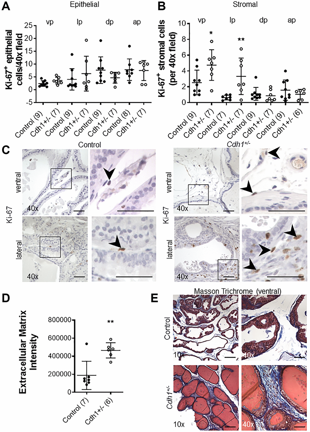
Figure 2.Impact of prostate epithelial specific heterozygous E-cadherin loss on proliferation and extracellular matrix deposition in the murine prostate at 24 months of age. (A) Quantification of Ki-67+ epithelial, and (B) stromal cells in the lobes of the prostate from Control and Cdh1+/- mice at 24 months of age. Abbreviations: vp: ventral prostate; lp: lateral prostate; dp: dorsal prostate; ap: anterior prostate. (C) Ki-67 immunostaining (brown) in the stromal compartments of prostate ventral and lateral lobes. Black arrows indicate Ki-67+ cells in the stromal compartment. Original magnification, 40×, inset 40×. (D) Quantification of Masson’s trichrome staining of extracellular matrix (blue) in the stroma surrounding prostate glands from ventral prostate of Control and Cdh1+/- mice at 24 months of age. Seven fields from each section were analyzed and an average score was determined for each mouse. (E) Masson’s trichrome staining in transverse sections of prostate ventral lobes in Control (top panels) and Cdh1+/- mice (bottom panels). Original magnification, 10×, inset 40×. Data represent mean ± S.D, number of mice in each group in parentheses. Lobes which had been washed away during staining process were not quantified. *p < 0.05, **p < 0.01. (D) Scale bars indicate 200 μm in 10×, 50 μm 40×.