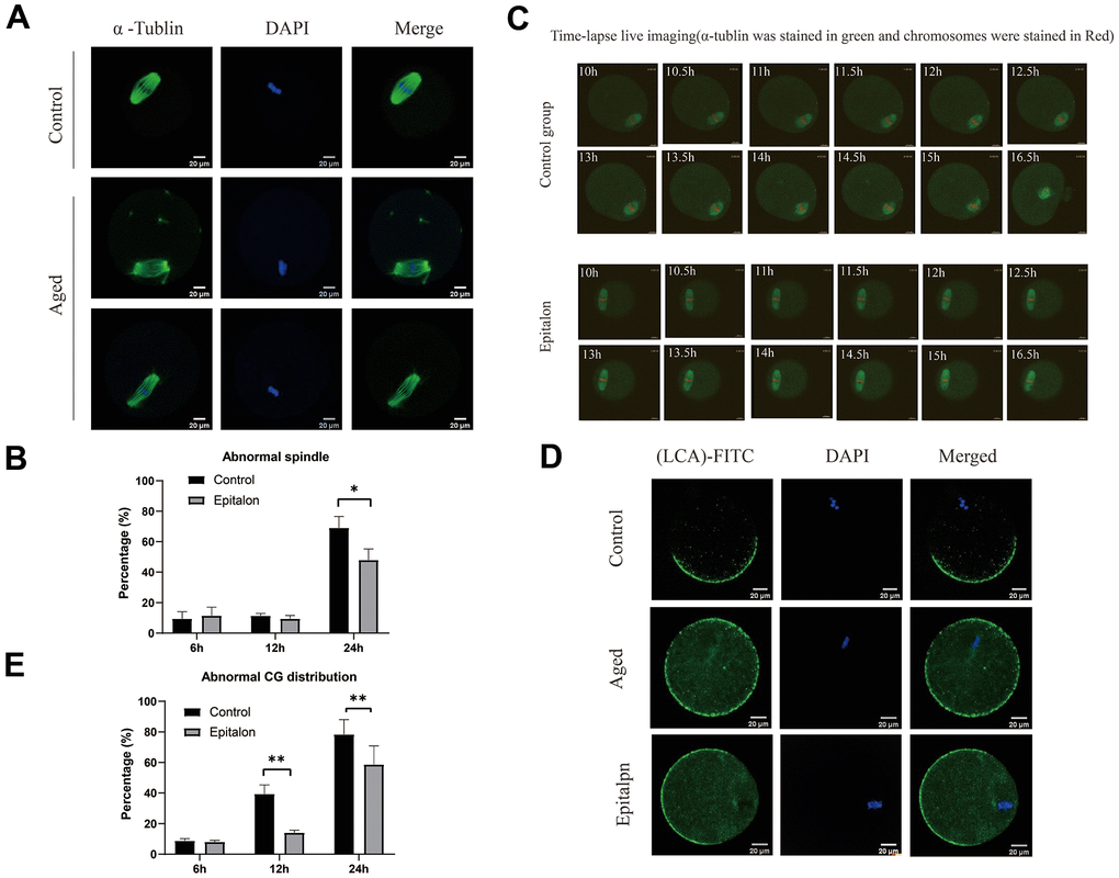
Figure 3.Epitalon maintained normal spindle integrity and CGs distribution. (A) Different morphological structures of spindles appeared in the Control and aged oocytes. Meiotic spindles in oocytes were stained with α-tubulin (green) and chromosomes were stained with Hoechst 33342 (blue). (B) Percentages of abnormal spindles in oocytes. Oocytes containing normal spindles and those with abnormal spindles were counted to calculate the percentage of abnormal spindles. (C) Dynamic spindle and chromosome changes in the Control and Epitalon treated oocytes, which were microinjected with MAP4-eGFP mRNA and H2B-mCherry mRNA, and visualized by time-lapse live-cell imaging. The spindles were marked in green, and chromosomes were marked in red. Independent replicates were conducted with a minimum of 20 oocytes. (D) Confocal laser scanning microscopy for the distributions of cortical granules (CGs) of oocytes. Metaphase II oocytes were immunolabeled for CGs with lens culinaris (LCA)-FITC (fluorescein isothiocyanate: green), and chromosomes were counterstained with DAPI (blue). (E) Percentages of abnormal distribution of CGs in oocytes. Significant difference between the Aged and 0.1mM Epitalon-treated groups was observed (p < 0.05). Data are expressed as mean ± SEM of at least three independent experiments. Scale bar: 20 μm.