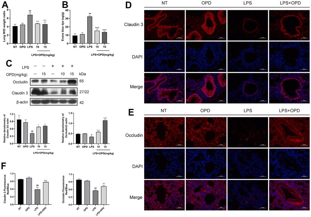
Figure 2.Oxypeucedanin maintains the integrity of the lung air-blood barrier in LPS-induced mice. LPS solution was dripped into one nostril of each mouse to establish the ALI model. Twelve hours later, a sample of mouse lung tissue was collected. OPD (10 and 15 mg/kg) was administered by intraperitoneal injection 1 h before the model was constructed. (A) The ratio of wet weight to dry weight of lung tissue. The lung wet/dry weight ratio was determined by the aforementioned method (n=5). (B) The mice were administered a tail vein injection of 1% Evans Blue (40 mg/kg, Sigma–Aldrich, MO, USA) 2 h before euthanasia. The measurement was then performed according to the method mentioned earlier. Pulmonary vascular permeability was observed by the increase in Evans blue dye in the lung tissue (n=5). (C) Protein levels of Occludin and Claudin 3 in LPS-treated mice (n=3). Quantitative analysis of Occludin and Claudin 3 proteins by ImageJ. (D–F) Immunofluorescence of Occludin and Claudin 3 proteins in lung tissue. Red indicates Occludin and Claudin 3 proteins, and blue indicates DAPI. Scale bar = 50 μm (n=3). Quantitative analysis of fluorescent pictures of Occludin and Claudin 3 by ImageJ. The concentration of the OPD animal experiment was 15mg/kg. SEM was used as the error standard for data analysis, and the experiment was repeated three times independently. #p < 0.01 and ##p < 0.0001 compared with No-treatment group; *p < 0.05, **p < 0.01 and ****p < 0.0001 compared with the LPS group. LPS: Lipopolysaccharide; OPD: Oxypeucedanin; W/D: Wet/Dry weight.