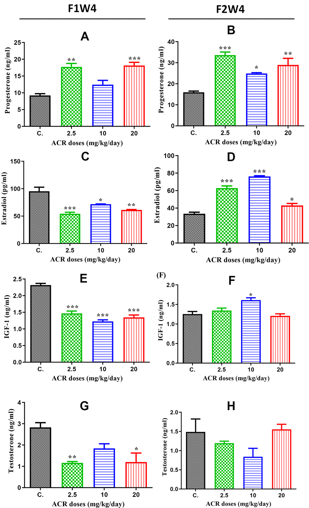
Figure 3.Hormone levels in the plasma of female offspring rats exposed to ACR during their fetal life compared to those in controls. (A) Plasma progesterone levels (ng/mL) in AF1 significantly increased in 2.5 and 20 mg/kg/day doses compared to the control group, while the increase in 10 mg/kg/day dose was nonsignificant. (B) Plasma progesterone levels (ng/mL) in AF2 were significantly elevated in all ACR dose (2.5, 10, and 20 mg/kg/day) groups compared to that in the control group CF2. (C) Plasma estradiol levels in AF1 groups recorded a high significant decrease in all doses of ACR. (D) A Plasma estradiol levels significantly increased in all AF2 treated groups compared to the control group CF2. (E) Plasma IGF-1 levels significantly decreased in all treated groups of AF1 compared to the control group. (F) We found a significant increase in plasma IGF-1 levels in the AF2 group treated with 10 mg/kg/day, whereas no significant variation was found in 2.5 and 20 mg/kg/day groups compared to the control group CF2. (G) Plasma testosterone levels in AF1 groups show a significant decline in 2.5 mg/kg/day and 20 mg/kg/day, whereas no significant variation was noted in 10 mg/kg/day. (H) No significant changes were observed in plasma testosterone levels in all AF2 treated groups compared to the control group CF2.