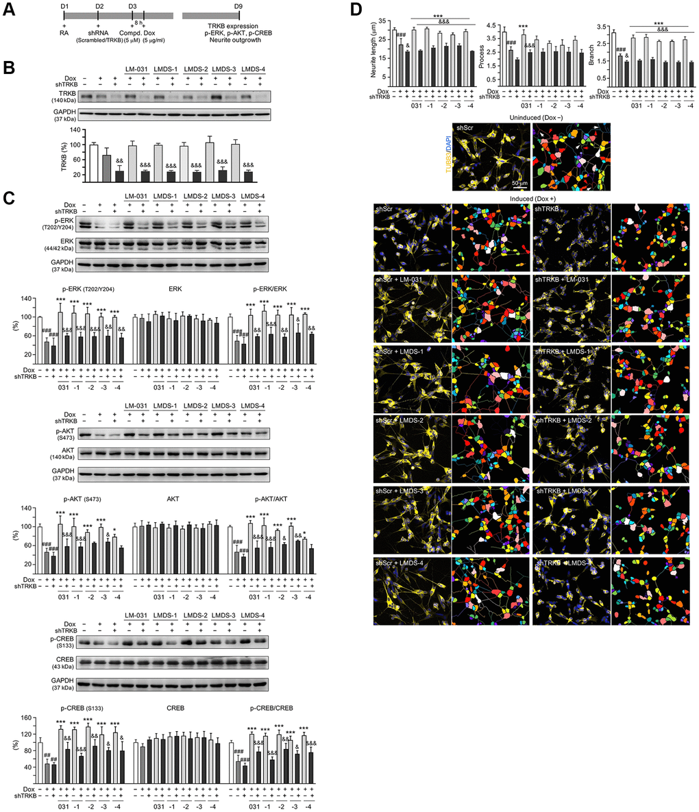
Figure 5.TRKB RNA interference of Aβ-GFP SH-SY5Y cells. (A) Experimental flow chart. On day 1, Aβ-GFP SH-SY5Y cells were plated with retinoic acid (RA; 10 μM). On day 2, the cells were infected with lentivirus-expressing TRKB-specific or scrambled shRNA. At 24 h post-infection, LM-031 or LMDS-1 to -4 (5 μM) was added to the cells for 8 h, followed by induction of Aβ-GFP expression (Dox, 5 μg/ml) for 6 days. On day 9, TRKB and neurite outgrowth analyses were performed. Western blot analysis of (B) TRKB, (C) p-ERK (T202/Y204), ERK, p-AKT (S473), AKT, p-CREB (S133), and CREB in compound-treated cells infected with TRKB-specific or scrambled shRNA-expressing lentivirus (n = 3). GAPDH was used as a loading control. To normalize, the relative protein level of uninduced cells was set at 100%. (D) Microscopic images and neurite outgrowth (length, process and branch) assay of Aβ-GFP-expressing cells with TRKB-specific or scrambled shRNA, and with or without LM-031 or analogs (5 μM) treatments (n = 3). TUBB3 staining (yellow) was used to quantify the extent of neurite outgrowth. Nuclei were counterstained with DAPI (blue). Also shown were segmented images with multi-colored mask to assign each outgrowth to a cell body for quantification. In uninduced cells, processes and branches are indicated with red and white arrows, respectively. P values: comparisons between induced vs. uninduced cells (###P < 0.001), compound-treated vs. untreated (induced) cells (***P < 0.001), or TRKB shRNA-treated vs. scrambled shRNA-treated cells (&P < 0.05, &&P < 0.01, &&&P < 0.001). (one-way ANOVA with a post hoc Tukey test).