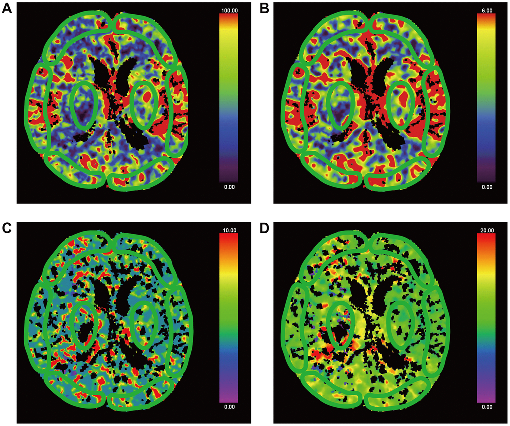
Figure 3.False-colour pictures of a 76-year-old MCS− man after right thalamus hemorrhage at the basal ganglia level. The affected side is right. The damaged part is right thalamus. (A) The CBF map of the bilateral frontal cortex, temporal cortex, occipital cortex and thalamus, (B) the CBV map, (C) the TTP map and (D) the MTT map.