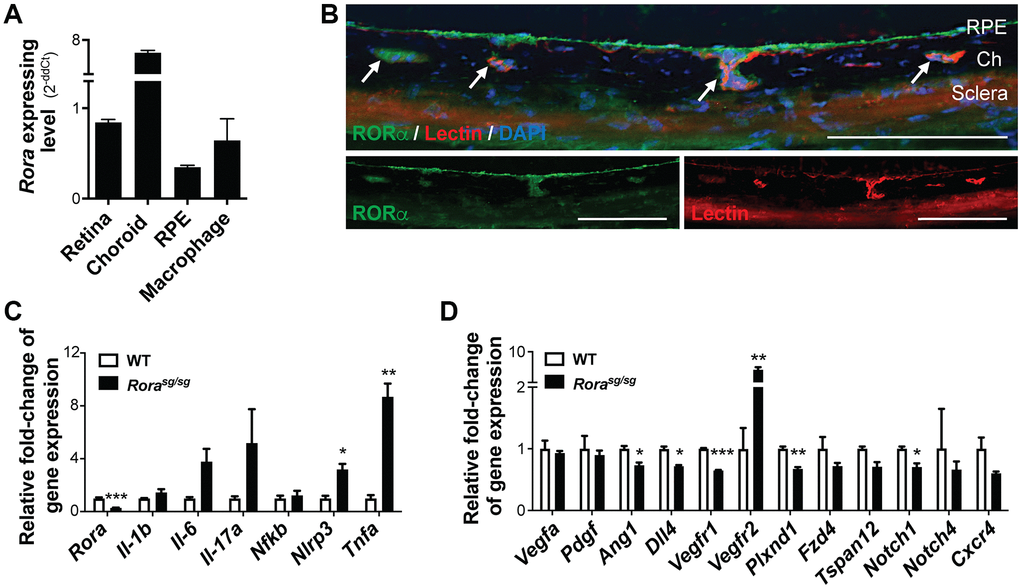
Figure 1.RORα was enriched in mouse choroid and regulated inflammatory and angiogenic genes. (A) Relative Rora expression in different types of mouse ocular tissues, namely retina and RPE/choroid complex, and cells (isolated pure RPE from mouse eyes and RAW264.7 macrophage cell line) measured with quantitative RT-PCR and normalized to housekeeping gene Rn18s. The choroid complexes expressed the highest expression levels of Rora compared to the retinas, RPE, and macrophage cells (n = 3/group). (B) Immunohistochemistry staining of retinal cross sections shows RORα antibody staining (green), vascular endothelium marker isolection (red), and DAPI (blue). Ch: choroid. Scale bars, 100 μm. (C, D) q-PCR analysis for the expression of Rora and inflammatory (C) and angiogenic (D) genes in the RPE/choroid complexes from Rorasg/sg and WT mice in normal condition without CNV showed that deficiency of RORα led to significant increase in Vegfr2 and Tnfa mRNA levels, in addition to changes in other inflammatory and angiogenic genes (n = 3 mice/group). Data are presented as means ± SEM. *P ≤ 0.05; **P ≤ 0.01; ***P ≤ 0.001.