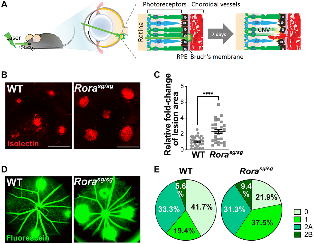
Figure 2.Genetic deficiency of RORα increased lesion size and vascular leakage in a mouse model of laser-induced choroidal neovascularization (CNV). (A) A cartoon illustrating laser-induced CNV model in mice. Young adult mice are exposed to laser, which ruptures Bruch’s membrane and causes CNV. (B) Representative images of choroidal flat mounts with laser-induced CNV from wild type (WT) and RORα-deficient (Rorasg/sg) mice stained with isolectin IB4 (red) showing four lesions, with optic disc in the center. Scale bars, 500 μm. (C) Quantification of the relative fold-change of CNV lesion areas showed that RORα-deficient mice have larger CNV lesion sizes compared to age-matched WT (n = 22–23 eyes/group). Each data point represents averaged lesion size from one eye. Solid horizontal bars indicate means ± SEM; ****P ≤ 0.0001. (D) Representative images of fundus fluorescein angiography (FFA) from WT and Rorasg/sg mice with laser-induced CNV at day 6 after laser photocoagulation. (E) Lesions were graded on an ordinal scale of the fluorescein (D; green) leakage appearance: grade 0 (no leakage); grade 1 (questionable leakage); grade 2A (leaky); grade 2B (pathologically significant leakage). Rorasg/sg mice revealed much fewer grade 0 lesions and more grade 1, 2A and 2B lesions compared to WT mice (n = 10 mice/group).