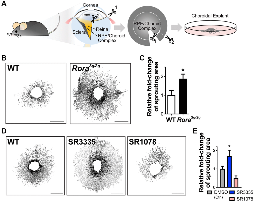
Figure 4.RORα regulates choroidal sprouting ex vivo. (A) A cartoon illustrates the experimental steps of choroidal explant assay by isolation, dissection and culture of choroid fragments. (B) Representative images of choroidal sprouting assays from age-matched WT and Rorasg/sg mice. Scale bars, 1 mm. (C) Quantitative analysis of the choroidal sprouting area from 5 days after explantation showed that Rorasg/sg choroids have significantly increased sprouting ability ex vivo compared to WT. n = 3–5 mice (10–12 explants)/group. (D) Representative images of choroidal explants isolated from C57BL/6J mice and treated with SR3335 (RORα inverse agonist), SR1078 (RORα/γ agonist) or vehicle control DMSO (all at 5 μM). Scale bars, 1 mm. (E) Quantification of the sprouting area indicated that inhibition of RORα with SR3335 significantly increased choroidal sprouting area while SR1078 reduced the choroidal sprouting ability compared to the DMSO (control) treated group. n = 3 mice/group; 10–12 explants per treatment. Data are presented as mean ± SEM. *P ≤ 0.05.