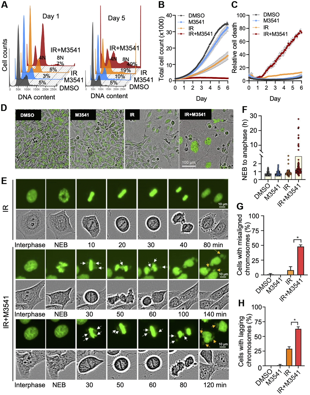
Figure 2.M3541 enhances radiation-induced cancer cells death by disrupting mitosis. (A) Cell cycle analysis of 7-AAD labeled HeLa cells exposed to DMSO, M3541, IR or IR+M3541 for 24 h and 5 days. Percentage of cells with more than 4N DNA content were calculated and shown from representative experiments. (B) Cell growth and (C) relative cell death in proliferating HeLa NucLight Green cells exposed to DMSO, M3541, IR or combination of IR+M3541 in the presence of Cytotox Red reagent. The cells were imaged every 2 h for 5 days by IncuCyte. Growth curves were built from the number of green-fluorescent nuclei at each time point. Relative cell death was determined as Cytotox Red positive counts normalized to green nuclei counts. Data are shown as mean ± SEM. (D) Representative still images from (B) were extracted from time-lapse videos taken by IncuCyte and shown at Day 5 post IR exposure. (E) Live cell imaging of HeLa cells expressing GFP tagged H2B exposed to IR or IR+M3541 by IncuCyte with a 20X objective. Individual cells were tracked in time-lapse videos and analyzed by ImageJ. Representative phase contrast and GFP images are shown. White arrows indicate chromosomal material that fails to align at the metaphase plate and lagging chromosomes. Orange arrows point to lagging chromosomes. (F) The lengths of time from nuclear envelop breakdown (NEB) to anaphase onset in HeLa GFP-H2B determined as in (E). (G) The percentage of metaphase cells with misaligned chromosomes and (H) the percentage of anaphase cells with lagging chromosomes were determined from time-lapse live imaging videos as in (E) Total 100 mitotic cells per condition were tracked from 2 independent experiments.