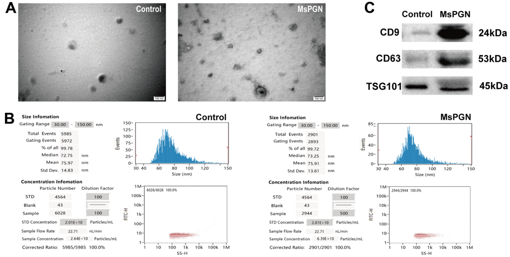
Figure 1.(A) Electron microscopic images of extracted exosomes revealed cup-shaped structures with a diameter of about 30–160 nm. Scale bar: 100 nm. (B) Nanoparticle tracking analysis (NTA) revealed the diameter of isolated extracellular vesicles (EVs) is consistent with that of exosomes. (C) Western blotting revealed CD9, CD63 and TSG101 proteins in exosome samples.