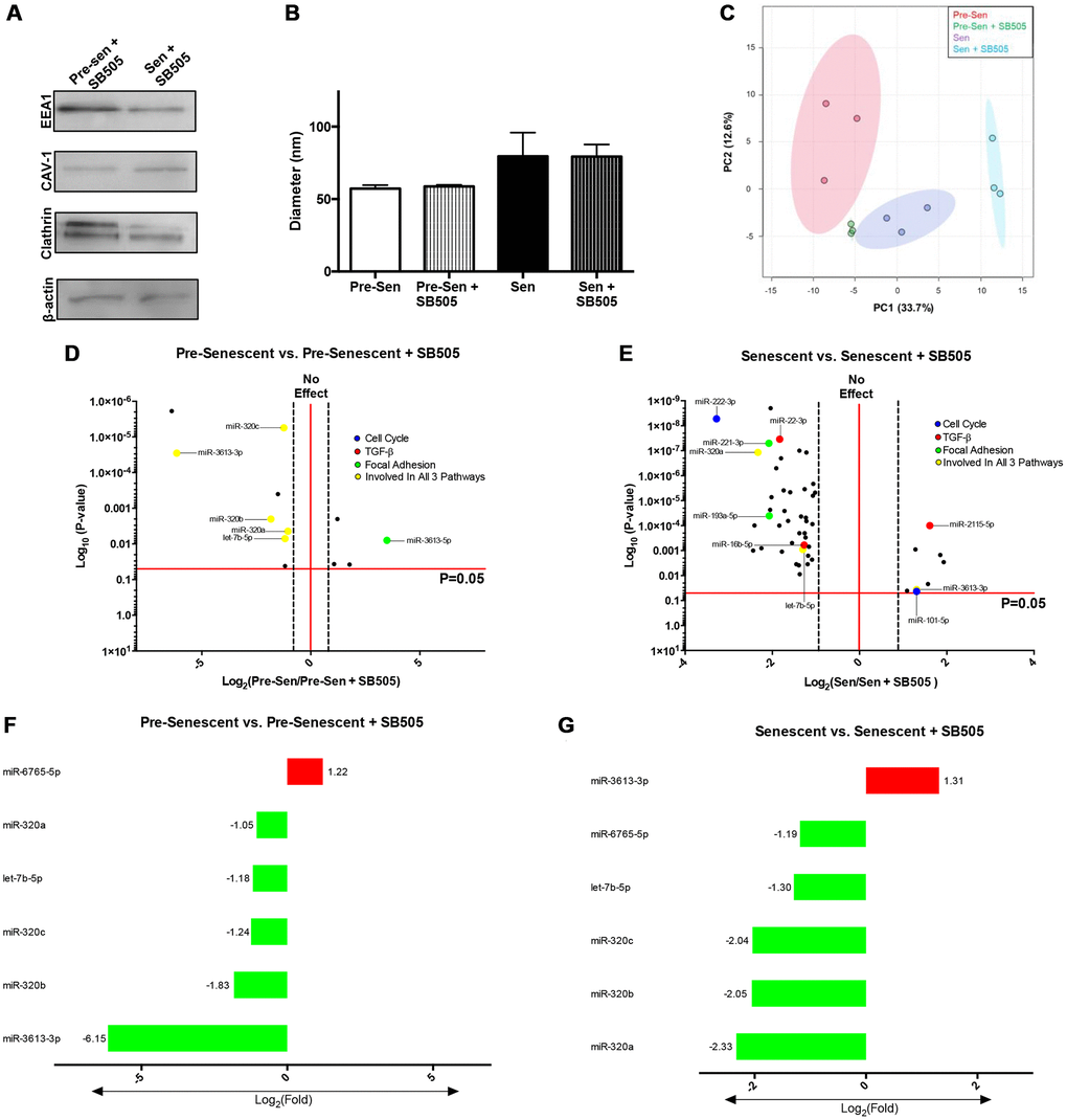
Figure 5.TGF-β modulates EXO miRNA profiles in pre- and senescent MSCs. EXO surface markers and miRNA content from pre- and senescent MSCs treated with SB505 were analyzed using immunoblots and microarrays. EXO populations were validated using immunoblots probing for well-known surface markers (A). Diameters were measured using dynamic light scattering methods and indicated similar diameters between untreated and SB505-treated EXOs from pre- and senescent MSCs; however, senescent MSC EXOs were larger than EXOs from pre-senescent cells (B). Principal component analysis (PCA) for pre- and senescent MSC EXOs treated with and without SB505 were plotted (pre-sen: red, pre-sen + SB505: green, sen: purple, sen + SB505: blue). Each point indicates a biological sample (n = 3) and the first two principal components explain 46.3% of miRNA variance (C). DIANA miRPath analysis showed that many of the significantly regulated miRNAs were involved in pathways of interest: cell cycle (blue), TGF-β (red), focal adhesion (green), and regulated in all 3 pathways (yellow) (D, E). 6 miRNAs overlapped between pre-senescent vs. pre-senescent + SB505 (F) and senescent vs. senescent + SB505 (G). In particular, expression levels between miRNAs-6765-5p and -3613-3p were inversely related between conditions. miRNA microarray samples were processed (n = 3) and miRNAs were considered significant if p < 0.05 and fold changes are reported as log2 differences.