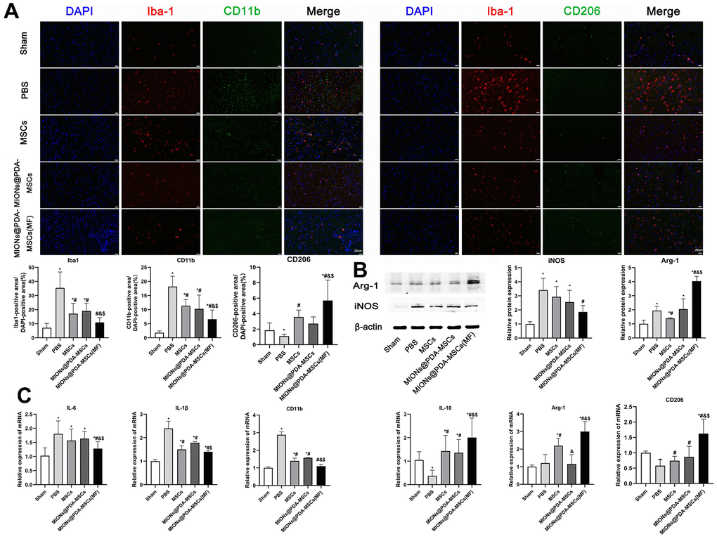
Figure 6.In vivo anti-inflammatory effects of the HUMSCs and MIONs@PDA-MSCs. (A) Immunofluorescence analysis and quantification for M1 (CD11b) and M2 (CD206) macrophage markers in the cortex tissues of mice. scale bars=20 μm. (B) Western blot analysis and quantification of M1 (iNOS) and M2 (Arg-1) macrophage markers in the cortex tissues of mice. (C) Relative expressions of M1 (IL-6, IL-1β and CD11b) and M2 (ARG-1, IL-10 and CD206) genes in the brain after treatment. *p < 0.05 vs. Sham group, #p <0 .05 vs. PBS group, &p <0 .05 vs. MSCs group, $p <0 .05 vs. MIONs@PDA-MSCs group.