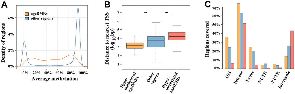
Figure 1.Region characteristics. (A) Distribution of methylation levels of ageDMRs versus other (non-significant) regions. The average methylation levels of ageDMRs (orange line) are predominantly in the mid-range (20-80%), whereas other regions (blue line) are either in the low range (< 20%) or high range (> 80%) of methylation. (B) Box plots showing the distance of analyzed regions to the nearest transcription start site (TSS). The median is represented by a horizontal line. The bottom of the box indicates the 25th percentile, the top the 75th percentile. The blue box represents non-significant regions, the orange box ageDMRs which lose methylation with age and the red box ageDMRs which gain methylation with age. Please note that hypomethylated ageDMRs are significantly (*** P < 0.001) closer to the TSS, whereas hypermethylated DMRs are more distant from the TSS than other regions. (C) Localization of hypomethlyated ageDMRs (orange bars) and hypermethylated DMRs (red bars) in different genic and intergenic regions, compared to non-significant regions (blue bars). Please note that some regions may be assigned to several gene parts and, therefore, the percentages of all bars (of one color) total > 100%.