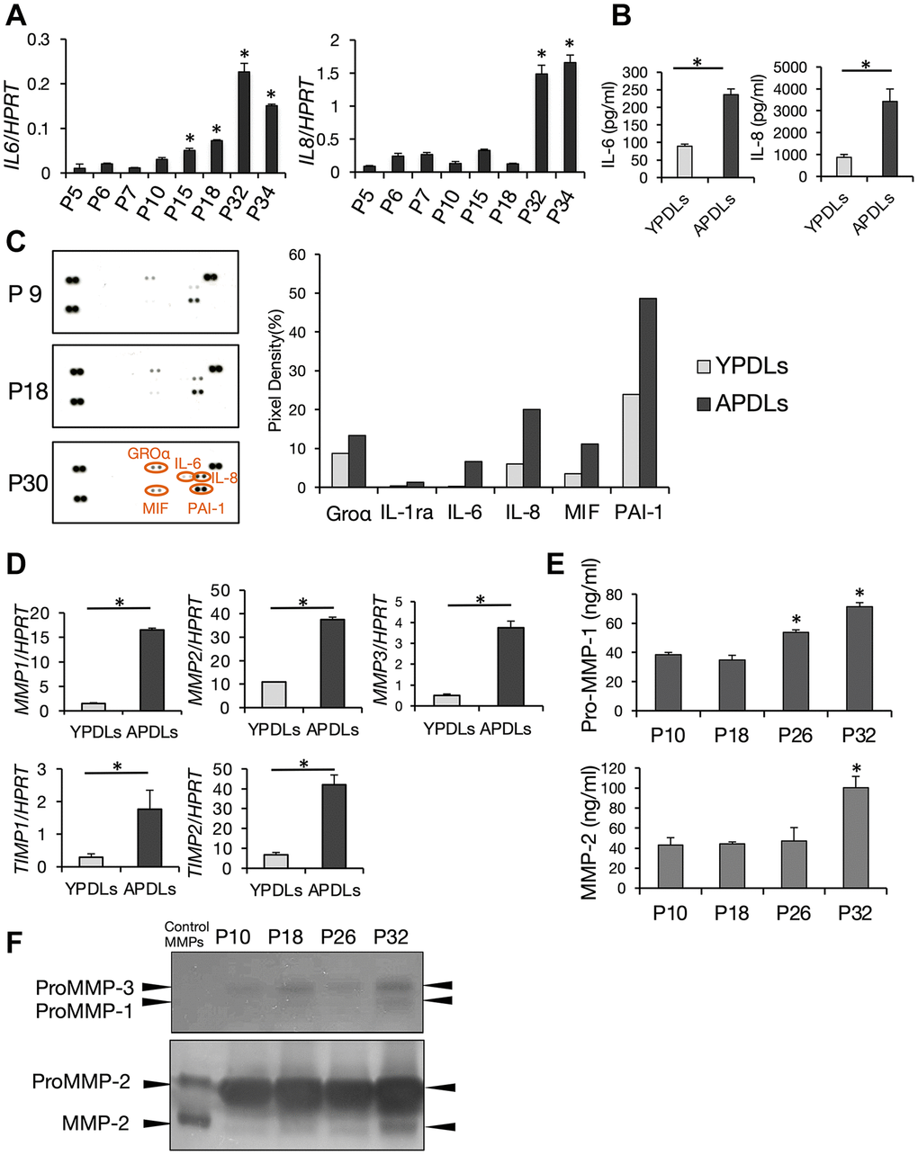
Figure 3.Increased expression of IL-6 and IL-8 in senescent HPDL cells. (A) Relative mRNA expression of IL-6 and IL-8 in various passages of HPDL cells quantified by qRT-PCR (*p < 0.01 vs. P5). (B) IL-6 and IL-8 in conditioned medium in YPDLs and APDLs (*p < 0.01). (C) Enhanced production of SASP factors in senescent HPDL cells. Soluble factors secreted by P9, P18, and P30 HPDL cells were detected by an antibody dot blot array. In right panels, quantification of signal intensity of dots plots assay for conditioned medium of YPDLs and APDLs. Signal intensities of the major dot blots were normalized against control spots in each blot and shown as bar graphs (Groa, IL-1ra, IL-6, IL-8, MIF, PAI-1). Gray bars indicate YPDLs (P9) and black bars indicate APDLs (P30). Representative data from three experiments are shown. (D) Relative mRNA expression of MMP-1–3 and TIMP-1 and -2 in HPDL cells quantified by qRT-PCR (*p < 0.01). (E) Pro-MMP-1 and MMP-2 in conditioned medium of P10, P18, P26, and P32 HPDL cells (*p < 0.01 vs. P10). (F) Inverted images of zymography for conditioned medium of P10, P18, P26, and P32 HPDL cells. Dark spots indicate Pro-MMP-1–3 and MMP-2. Representative data from three experiments are shown.