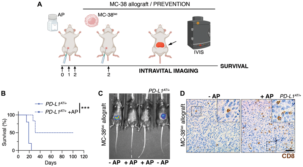
Figure 5.Depleting PD-L1+ cells prolongs survival in an immunocompetent model of peritoneal cancer metastasis. (A) Schematic overview of the prevention experimental workflow. 5 × 10⁵ MC-38luc cells were intraperitoneally injected into mice that were previously injected i.p. for 3 consecutive days with AP20187 (2.5 mg/kg). (B) Kaplan-Meier survival curve of control and AP20187-pretreated PD-L1AT/+ mice after i.p. inoculation of MC-38luc allografts. The p value was calculated with the Mantel-Cox log rank test. ***p < 0.001. (C) Representative IVIS image of mice from the experiment defined in (B) at day 4 post-tumor injection. (D) IHC of CD8 in intraperitoneal MC-38luc allografts isolated from control and AP20187-treated PD-L1AT/+ mice. Insets in each panel are magnified to illustrate the presence of tumor-infiltrating CD8+ cells. Scale bar (black) indicates 30 μm.