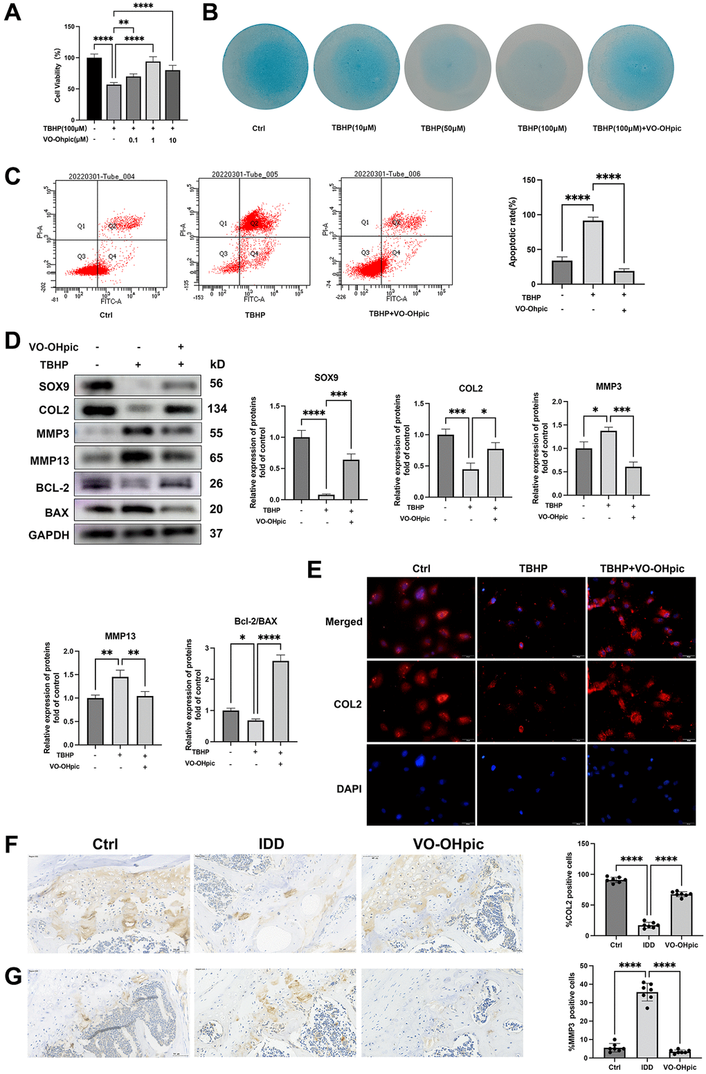
Figure 2.VO-OHpic inhibited oxidative stress induced endplate chondrocytes apoptosis and CEP degeneration. (A) CEP chondrocytes were isolated and treated with 100 μM TBHP with increasing concentrations (0, 0.1, 1, 10 μM) of VO-OHpic for 24 hours, CCK assay was conducted to evaluate the cell viability. (B) CEP chondrocytes were treated with increasing concentrations of TBHP and 1 μM VO-OHpic for 7 days and Alcian blue staining was conducted to examine the ECM production. (C) Flow cytometric analysis of endplate chondrocytes stained with Annexin V-FITC/PI. Percentage of apoptosis rates was expressed as means ± SD from three independent experiments. (D) CEP chondrocytes were pretreated with VO-OHpic (1 μM) for 18 hours, then 100 μM TBHP was added for 6 hours. Western blot was conducted to examine the protein levels of SOX9, COL2, MMP3, MMP13, BCL-2 and BAX. The band density of SOX9, COL2, MMP3, MMP13 and the ratio of BCL-2/BAX were quantified and normalized to control. (E) CEP chondrocytes were pretreated with VO-OHpic (1 μM) for 18 hours, then 100 μM TBHP was added for 6 hours. Immunofluorescence staining was conducted to examine the expression of COL2 (red). Scale bar = 20 μm. (F, G) Immunohistochemistry for COL2 and MMP3 in cartilage endplate from each group. Scale bar = 20 μm. The ratio of positive cells for COL2 and MMP3 was quantified under a microscope at 400× magnification using five sections from seven mice. Data are presented as mean ± SD from three independent experiments. *P < 0.05, **P < 0.01, ***P < 0.001, ****P < 0.0001.