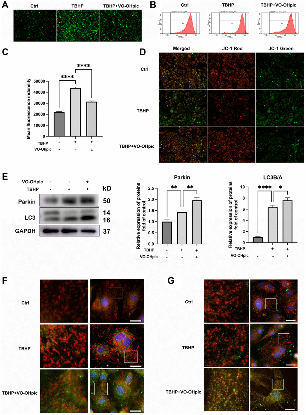
Figure 4.VO-OHpic treatment promoted mitophagy in CEP chondrocytes. (A) CEP chondrocytes were treated with TBHP (100 μM) and VO-OHpic (1 μM) for 24 h, representative fluorescence microscopy photomicrographs of intracellular ROS in chondrocytes. (B) ROS was detected by flow cytometric analysis after labeling with DCFH-DA. (C) The bar graphs show the mean fluorescence intensity of ROS levels in endplate chondrocytes. (D) Representative fluorescence microscopy photomicrographs of mitochondrial membrane potential (MMP) after incubating with JC-1. Red fluorescence was emitted by JC-1 aggregates in healthy mitochondria with polarized inner mitochondrial membranes, whereas green fluorescence was emitted by cytosolic JC-1 monomers, indicating MMP collapse. (E) CEP chondrocytes were pretreated with VO-OHpic (1 μM) for 18 hours, then 100 μM TBHP was added for 6 hours, western blot was conducted to examine the protein levels of parkin and LC3. The band density of parkin and LC3 was quantified and normalized to control. (F, G) Immunofluorescence staining was conducted to examine the expression and localization of LC3B, parkin (green) and mitochondria (red). Scale bar = 25 μm. Data are presented as mean ± SD from three independent experiments. *P < 0.05, **P < 0.01, ****P < 0.0001.