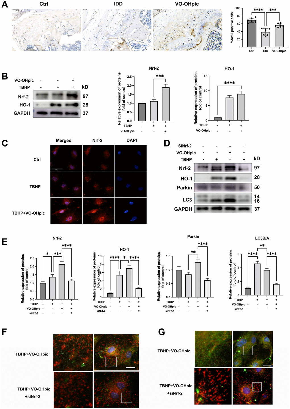
Figure 6.Nrf-2 activation is required for VO-OHpic induced mitophagy process. (A) Immunohistochemistry for Nrf-2 in cartilage endplate from Ctrl group, IDD group and IDD+VO-OHpic group. Scale bar = 50 μm. The ratio of positive cells for COL10 was quantified under a microscope at 400× magnification using five sections from seven mice. (B) CEP chondrocytes were pretreated with VO-OHpic (1 μM) for 18 hours, then 100 μM TBHP was added for 6 hours. Western blot was conducted to examine the protein levels of Nrf-2 and HO-1. The band density of Nrf-2 and HO-1 was quantified and normalized to control. (C) CEP chondrocytes were treated with TBHP (100 μM) and VO-OHpic (1 μM) for 24 h and immunofluorescence staining was conducted to examine the expression and localization of Nrf-2 (red). Scale bar = 20 μm. (D) Chondrocytes were transfected with Nrf-2 siRNA, and treated with TBHP (100 μM) and VO-OHpic (1 μM), western blot was conducted to examine the protein levels of Nrf-2, HO-1, parkin and LC3. (E) The band density of Nrf-2, HO-1, parkin and LC3 was quantified and normalized to control. (F, G) Chondrocytes were transfected with Nrf-2 siRNA, and treated with TBHP (100 μM) and VO-OHpic (1 μM). Immunofluorescence staining was conducted to examine the expression and localization of LC3B, parkin (green) and mitochondria (red). Scale bar = 25 μm. Data are presented as mean ± SD from three independent experiments. *P < 0.05, **P < 0.01, ***P < 0.001, ****P < 0.0001.