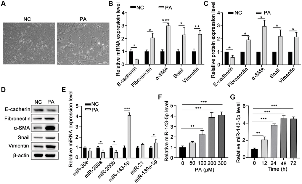
Figure 1.PA induced EMT and increased miR-143-5p expression in ARPE-19 cells. (A) Morphological alteration of ARPE-19 cells after treating with PA. Scale bar, 200 μm. (B) mRNA expression of E-cadherin, Fibronectin, α-SMA, Snail, and Vimentin as determined via quantitative PCR (qPCR) in ARPE-19 cells after PA treatment (n = 3 for each group). (C, D) The protein expression of E-cadherin, Fibronectin, α-SMA, Snail, and Vimentin determined using western blot in ARPE-19 cells after PA treatment (n = 3 for each group). (E) The expression of the indicated miRNAs in untreated control (NC) ARPE-19 cells, and ARPE-19 cells treated with PA (n = 3 for each group). (F) qPCR assays in dose-course experiments showed that the optimum PA concentration for inducing miR-143-5p expression in RPE cells was 200 μM (n = 5 for each group). (G) Results of time-course experiments demonstrated that the optimum duration of PA induction of miR-143-5p expression in RPE cells was 48 h (n = 5 for each group). Data are expressed as mean ± standard deviation; *P < 0.05, **P < 0.01, and ***P < 0.001, as compared with the indicated controls.