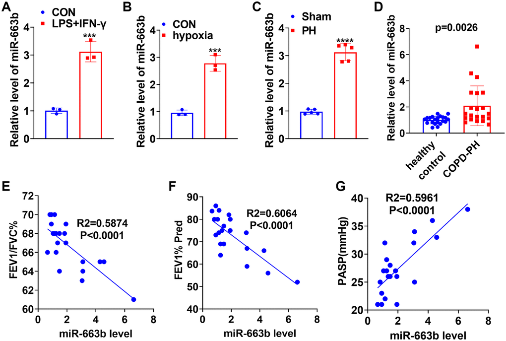
Figure 1.miR-663b presented a high expression in M1 macrophages as well as in the in-vivo and in-vitro pulmonary hypertension models. (A) LPS (10 μg/mL) plus IFN-γ (20 ng/ml) treated THP-1 cells for 24 hours to elicit macrophage M1 polarization. RT-PCR determined miR-663b expression in M1 macrophages. (B) Hypoxia-treated PASMCs in order to build an ex-vivo pulmonary hypertension model. RT-PCR confirmed miR-663b expression in PASMCs treated with hypoxia. N = 3. ***P < 0.001 (vs. CON). (C) A pulmonary hypertension rat model induced by hypoxia was constructed, with RT-PCR conducted to check miR-663b expression in the rat serum. (D) RT-PCR was conducted to detect miR-663b expression in the serum of COPD-PH patients (n = 22) and health controls (n = 22). (E–G). Pearson linear regression analysis was used for analyzing the associations of serum miR-663b level with FEFV1/FVC% (E), FEV1% Pred (F) and PASP (mmHg) (G). N = 5. ***P < 0.001, ****P < 0.0001 (vs. CON or Sham).