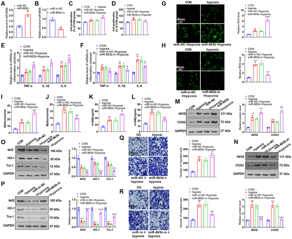
Figure 3.miR-663b overexpression facilitated PASMC damage induced by hypoxia. (A, B) PASMCs were transfected along with miR-NC, miR-663b mimics, miR-in, and miR-663b inhibitors, with RT-PCR implemented 48 hours later to confirm miR-663b expression in the transfected cells. Then, hypoxia was harnessed for 24-hour treatment of the transfected cells. (C, D) CCK8 assay was conducted for examining cell proliferation. (E, F) RT-PCR checked the levels of inflammatory cytokines TNF-α, IL-1β, and IL-6 in PASMCs. (G–L) Cell immunofluorescence and colorimetry determined the levels of ROS (G, H), MDA (I, J), and 4-HNE (K, L) in PASMCs. (M–P) Western blot was performed for assaying the profiles of inflammation-concerned proteins iNOS and COX2 and oxidative stress-correlated proteins Nrf2, HO-1 and Trx-1 in PASMCs. (Q, R) Transwell monitored PASMC migration. N = 3. *P < 0.05, **P < 0.01, ***P < 0.001 (vs. miR-NC, miR-in-NC, or CON); +P < 0.05, ++P < 0.01, +++P < 0.001 (vs. miR-NC+hypoxia or miR-in-NC+hypoxia).