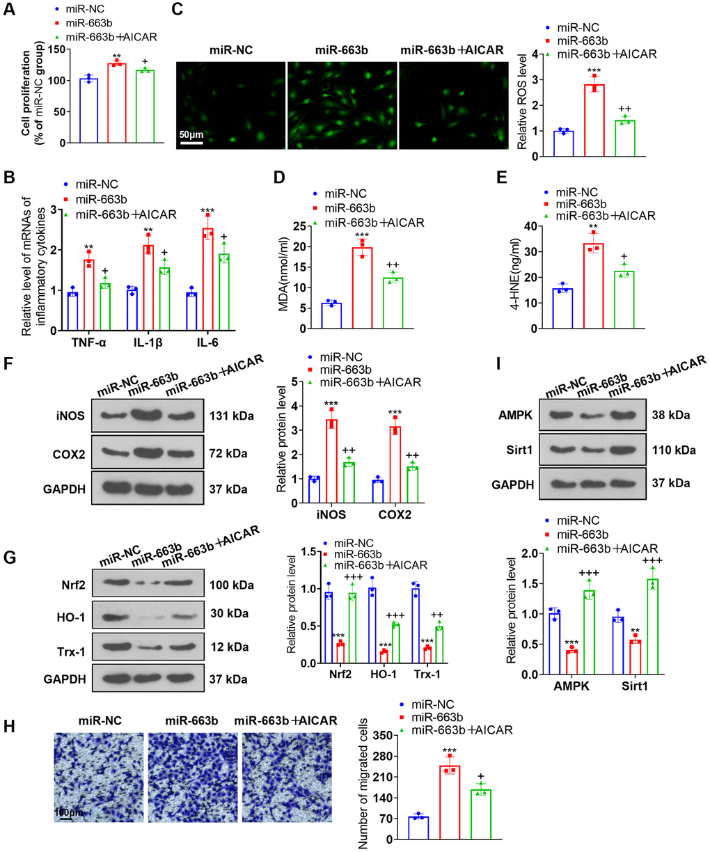
Figure 6.AMPK activation alleviated the effects of miR-663b overexpression on PASMCs. miR-663b mimics were transfected into PASMCs, with the AMPK activator AICAR applied for treatment. (A) CCK8 assay was conducted for examining cell proliferation. (B) RT-PCR measured TNF-α, IL-1β, and IL-6 profiles in PASMCs. (C) Cell immunofluorescence determined ROS levels in PASMCs. (D, E) Colorimetry confirmed MDA and 4-HNE contents in PASMCs. (F) Western blot verified iNOS and COX2 levels in PASMCs. (G) Western blot checked Nrf2, HO-1 and Trx-1 expressions in PASMCs. (H) Transwell monitored PASMC migration. (I) Western blot figured out the profiles of AMPK and Sirt1 in PASMCs. N = 3. **P < 0.01, ***P < 0.001 (vs. miR-NC); +P < 0.05, ++P < 0.01, +++P < 0.001 (vs. miR-663b).