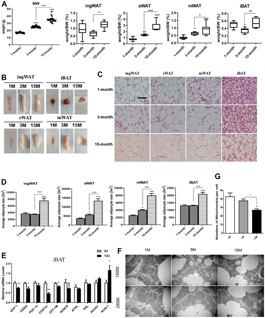
Figure 1.Differences in fat mass and fat distribution between young, adult and middle-age transitional mice. Young mice (1-month-old), adult mice (3-month-old) and middle-age transitional mice (15-month-old) were fed ad libitum. Body weight and body adipose tissues ratio of inguinal white adipose tissue (ingWAT), epididymal WAT (eWAT), mesenteric WAT (mWAT), interscapular BAT (iBAT) of mice (A) and their appearance (B) were analyzed (n = 6–12). The effect of aging on histological characteristics of white and brown adipose tissues (C). The average area of adipocytes (μm2) in every 100-mm2 area range of various adipose tissues was quantified using the Image Pro Plus software (D) (n = 6-8). Scale bar represents 100 μm. Differential expression of mitochondrial biogenesis related gene (Pgc-1α) and thermogenic gene (Ucp-1) (E) were measured (n = 6). Representative transmission electron microscopy (TEM) images of brown adipose tissues from the three groups of mice fed ad libitum and mitochondrial numbers (F, G) were analyzed. All data are presented as the mean ± SEM. * p < 0.05; ** p < 0.01; *** p < 0.001.