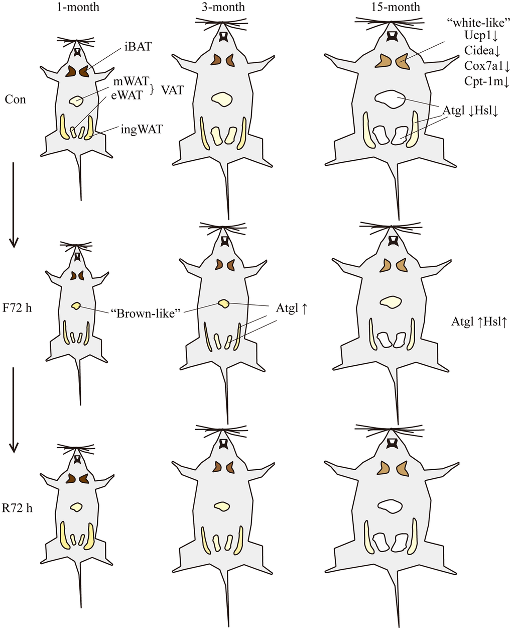
Figure 5.Schematic showing the response of mice of different ages to fasting and refeeding. With the increase of age, the body weight and BATR of all four fat depots increased. The brown adipocytes of the middle-aged transitional mice appeared “white-like”, and the mitochondrial thermogenesis gene Ucp-1, brown tissue marker genes Cidea and mitochondrial related genes Cox7a1 and Cpt-1m decreased; the expression of lipolytic genes Atgl and Hsl were lower relative to adult mice. F72 h significantly reduced the total white BATR (especially mesenteric adipose tissue) of the three groups of mice. Among them, the BATR of various adipose tissue depots of the middle-aged transitional mice showed the smallest decrease; and the expression level of Atgl in fat tissues of adult mice increased. After R72 h, the total white BATR of the three groups of mice was still significantly lower compared to the normal feeding group. Among them, the BATR of sWAT returned to normal, while the VAT was still low. The adipose tissue of middle-aged transitional mice reacted slowest to fasting and refeeding cues. Different size and color of adipose tissue represent different morphological changes induced by fasting or refeeding.