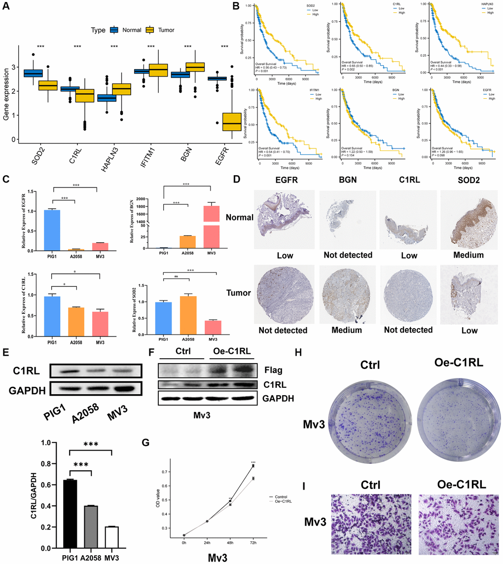
Figure 10.Prognostic TRGs gene expression levels in tumor and normal cells and functional analysis. (A) Different expressions of the six signature genes between the normal and tumor tissues (TCGA AND GTEx). (B) Confirmation of prognostic value of 6 TRGs for patients in TCGA by Kaplan–Meier analysis. (C) The relative RNA levels of EGFR, BGN, C1RL AND SOD2 in normal skin melanocyte and melanoma cell lines by q-PCR. (D) The IHC staining showed 4 signature genes expression at the protein level. (E) The protein level expression of C1RL genes based on melanocytes (PIG1) and melanoma cells lines (A2058 and MV3) by Western blot. (F) Western blot is used to assess the Overexpression efficiency of in MV3. CCK8 (G) and colony formation assay (H) are performed to assess effects of C1RL Overexpression on proliferation of MV3. (I) Transwell assay is utilized to evaluate the migration.