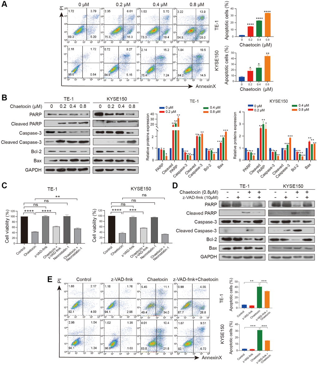
Figure 3.Chaetocin induces ESCC cells apoptosis in a caspase-dependent manner. (A) TE-1 and KYSE150 cells were treated with the indicated concentrations of chaetocin for 24 h, and apoptotic rates were detected using Annexin V/PI staining and flow cytometry. Results are shown as mean ± SD of three independent experiments. *P < 0.05, **P < 0.01, ***P < 0.001, ****P < 0.0001 vs. control group. (B) Western blot analysis of PARP, Cleaved PARP, caspase-3, cleaved-caspase-3, Bax and Bcl-2 following treatment with 0–0.8 μM chaetocin for 24 h. GAPDH was utilized as an internal standard. Blots presented here are representative of three independent experiments. *p < 0.05, **p < 0.01, ***p < 0.001 compared with the control group. (C) TE-1 and KYSE150 cells were pretreated with Z-VAD-FMK (10 μM, 2 h) or necrostatin-1 (20 μM, 2 h) before chaetocin treatment (0.8 μM, 24 h), and the cell viability was analyzed by CCK8 assay. (D) Expression levels of PARP, Cleaved PARP, caspase-3, cleaved-caspase-3, Bax and Bcl-2 were detected by western blot. GAPDH was used as the loading control. (E) Apoptosis was analyzed by flow cytometry. Results in (C) and (E) are shown as mean ± SD of three independent experiments. **P < 0.01, ***P < 0.001, ****P < 0.0001, ns nonsignificant compared with the control group.