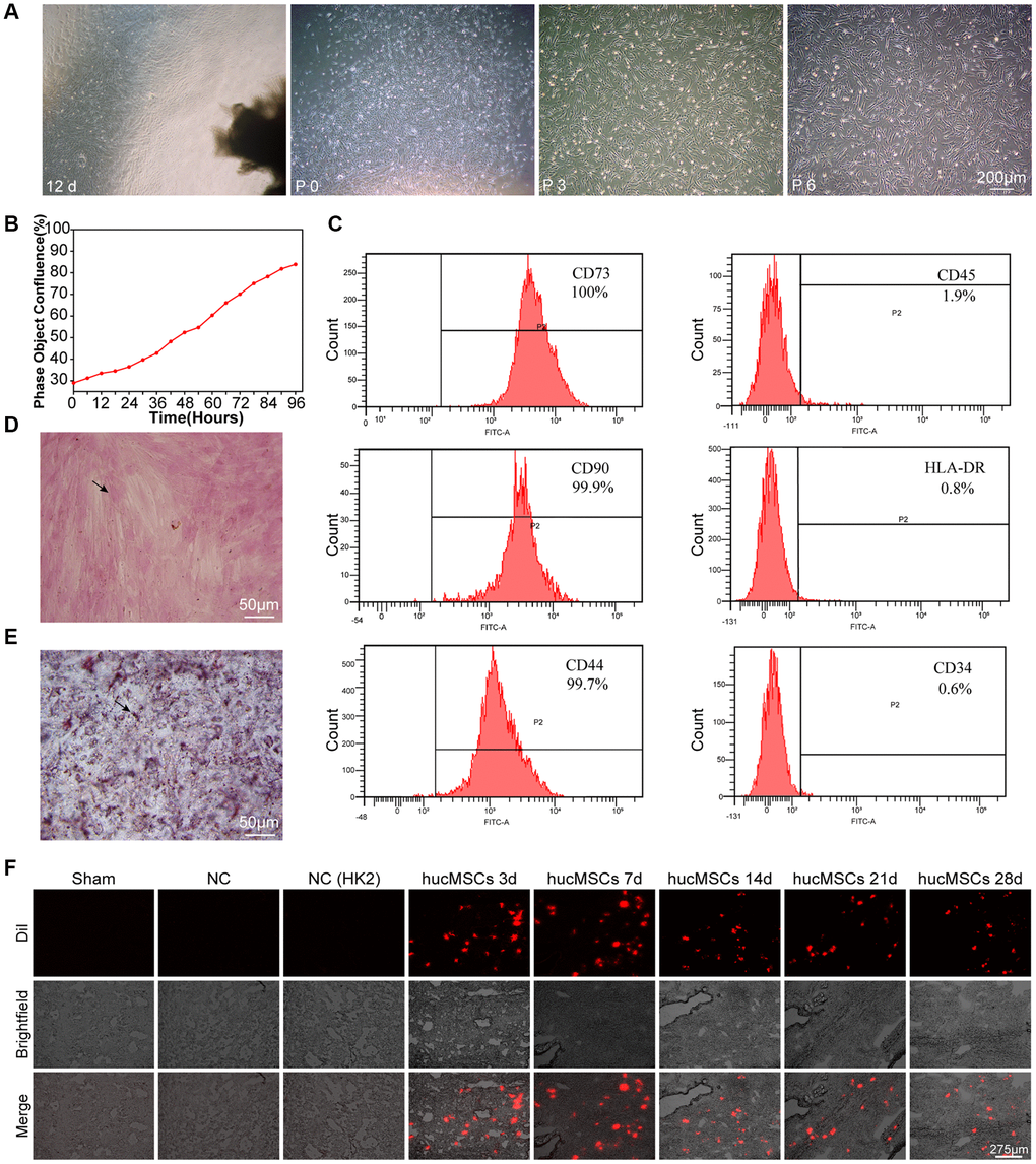
Figure 1.Culture and identification of hucMSCs. (A) Morphological observation of hucMSCs at different times. After 12 days of primary cultivation, cells developed around the tissue block. Zero generation of hucMSCs. Third generation of hucMSCs. Sixth generation of hucMSCs. (B) Growth curve of the third generation of hucMSCs showing their excellent growth as recorded by an IncuCyte S3 instrument. (C) Flow cytometry indicated that the positive rate was 100% for CD73, 99.90% for CD90, and 99.70% for CD44. The positive rate was 0.6% for CD34, 1.9% for CD45, and 0.8% for HLA-DR. (D) After 14 days of osteogenic induction, a positive reaction was noted between calcium nodus alizarin red stain and the osteogenic inducer. (E) After 28 days of adipogenic induction, fat droplets of verified size were positive for oil red O staining. (F) Images of cell membrane fluorescent probe revealing that hucMSCs were retained in the lungs. Mice injected with serum-free medium were used as sham. Mice injected with hucMSCs without fluorescent labeling were used as NC-1 to prove the absence of biological autofluorescence. Mice injected with human kidney 2 (HK-2) cells with labeled fluorescence were used as NC-2 to prove the absence of fluorescence dye leakage.