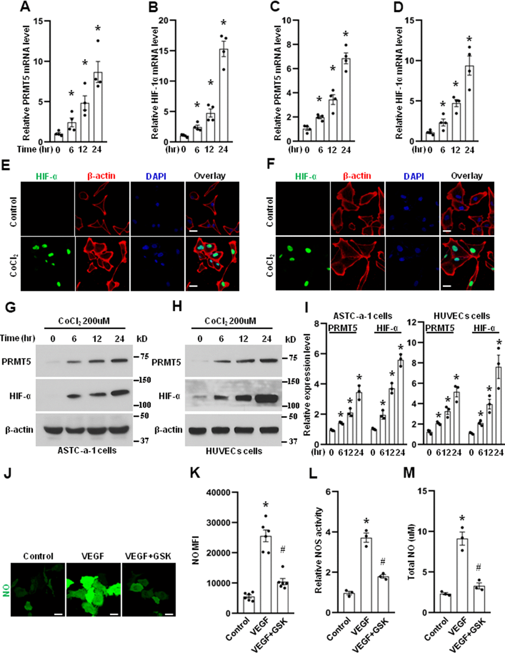
Figure 2.PRMT5 is induced by hypoxia. The HUVECs (A, B) and ASTC-a-1 cells (C, D) were treated with CoCl2 (200μM) for indicated time points, and the mRNA expression levels of PRMT5 and HIF-1α were measured by qRT-PCR. *P < 0.05 vs. 0-time point, n=4. The HUVECs (E) and ASTC-a-1 cells (F) were treated with or without CoCl2 (200μM) for 12h, and the HIF-1α expression induced by CoCl2 was detected by immunofluorescence staining. Representative pictures were shown. Green= HIF-1α; Red=β-actin; Blue=DAPI. Scale Bar=50μm. The ASTC-a-1 cells (G) and HUVECs (H) were treated with CoCl2 (200μM) for indicated time points, and the protein expression levels of PRMT5 and HIF-α were evaluated by Western blotting. (I) PRMT5 and HIF-α protein expression levels were quantified in ASTC-a-1 and HUVECs cells (n=3). *P < 0.05 vs. 0-time point. (J) The HUVECs were treated with VEGF (50ng/ml) in the presence or absence of GSK591, and the endothelial NO was monitored by the DAF-FM DA probe. Scale bar=50μm. (K) Quantitation of corresponding MFI values (n=6, each group). *P < 0.05 vs. control; #P < 0.05 vs. VEGF. (L) NOS enzymatic activity in HUVECs upon treatment of VEGF with or without GSK591 as evaluated by the Griess method. *P < 0.05 vs. control; #P < 0.05 vs. VEGF (50ng/ml) treatment. (n=3, each group). (M) Total NO was measured by ELISA kit using the Griess reaction in the supernatant of HUVECs upon treatment of VEGF (50ng/ml) with or without GSK591. *P < 0.05 vs. control; #P < 0.05 vs. VEGF treatment. (n=3, each group).