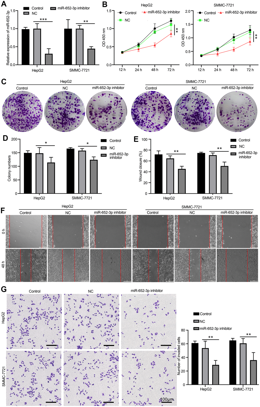
Figure 3.Inhibit miR-652-3p attenuates the proliferation and metastasis of HCC cells after co-culturing with BMSCs. (A) Expression of miR-652-3p was detected in HepG2 and SMMC-7721 cells co-culturing with BMSCs or hypo-BMSCs by using qPCR assay. Data were presented as the mean ± SD, and analyzed with Student’s t-test. **P < 0.01; *** P < 0.001, compared with the indicated controls. (B) Cell growth curves in HepG2 and SMMC-7721 cells transfected with different combinations. Data were presented as the mean ± SD, and analyzed with Student’s t-test. *P < 0.05; ** P < 0.01, compared with the indicated NC controls. (C) Colony formation assays showed that the proliferation rate was decreased in HepG2 and SMMC-7721 cells after treat isolated exosome hypo-BMSCs-derived with miR-652-3p inhibitor. (D) The numbers of colony were counted from six fields of view in each group. Data were presented as the mean ± SD, and analyzed with Student’s t-test. *P < 0.05. (E) The distance of migration was measured from six fields of view in each group. Data were presented as the mean ± SD, and analyzed with Student’s t-test. *P < 0.05; ** P < 0.01. (F) The numbers of dot violet were counted from six fields of view in each group. Data were presented as the mean ± SD, and analyzed with Student’s t-test. *P < 0.05; ** P < 0.01. (G) Cell invasion were measured by transwell assays. HepG2 and SMMC-7721 cells co-culturing with isolated exosome hypo-BMSCs-derived with miR-652-3p inhibitor treat for 48 h. Cells that invaded to the bottom surface were stained with crystal violet and observed by light microscopy (magnification, 100×).